
Cancer Cell Invasion – Marco Gerling's research group

We study how tumor cells invade healthy adjacent tissue. Our aim is to understand intercellular crosstalk in a way that is directly applicable to clinical practice. Our group consists of basic researchers and clinicians.

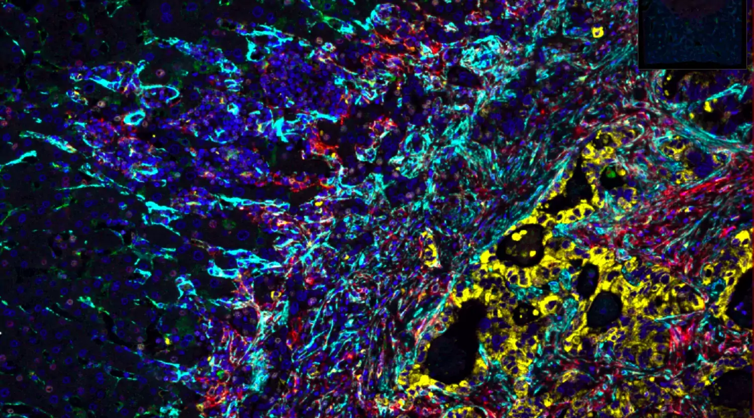
Fluorescent protein stains of tumor growing in the liver.

Colorectal and Pancreatic Cancer

Colorectal and pancreatic cancers account for more than 5,000 new cases every year in Sweden alone, and both frequently spread to the liver.

We study what happens where these tumors meet normal tissue. At this interface, cancer cells reshape their surroundings. These local interactions determine how tumors grow and invade, and they are reflected in distinct histological growth patterns that predict patient outcomes.

To chart these interactions, we combine spatial transcriptomics, digital pathology of whole-slide images, and experimental models. We validate our findings in large clinical cohorts at Karolinska University Hospital, including KaroLiver, a population-based, growing and living research resource of more than 800 patients treated for colorectal liver metastases, and in pancreatic cancer, where we map the tissue niches that protect tumor cells from therapy.

Our goal is to bridge clinical and basic research: to turn insights into tumour–tissue interactions into biomarkers and new treatment strategies, tested in clinical studies such as IMAGINE.
Histology of a liver metastasis
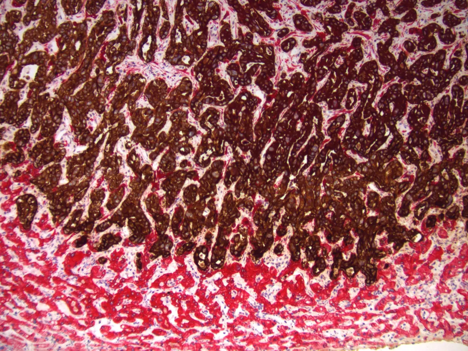
Histologic image of a liver metastasis, stained with antibodies for different cytokeratins. Tumor cells in brown, hepatocytes in red. Photo: Carlos F Moro

Liver Metastases

When tumor cells metastasise to distant organs, they encounter entirely new interaction partners.

Most metastases of gastrointestinal cancers are found in the liver, where tumor cells meet novel cell types, such as hepatocytes, hepatic stellate cells, liver sinusoid cells, and Kupffer cells. 

Little is known about tumor-microenvironment interactions in metastases, despite their clinical importance. Therefore, we have developed models to alter the metastatic microenvironment in the liver, and we analyse clinical samples from patients with liver metastases. Our aim is to understand how cancer cells progress in the liver parenchyma, in order to find ways to slow down or even revert this process.
Publications

All publications from group members

Selected publications

An idiosyncratic zonated stroma encapsulates desmoplastic liver metastases and originates from injured liver. Fernández Moro, Geyer, Harrizi, Hamidi et al. Nature Communications, https://doi.org/10.1038/s41467-023-40688-x

Analysis of the perimetastatic capsule that surround metastases with a particularly good prognosis.

Histopathological growth patterns of liver metastasis: updated consensus guidelines for pattern scoring, perspectives and recent mechanistic insights. Latacz, Höppener, Bohlok et al., British Journal of Cancer, https://doi.org/10.1038/s41416-022-01859-7

Comprehensive clinicopathological guidelines for growth pattern scoring of liver metastases with a dash of novel hypothesis.

Stabilization of the classical phenotype upon integration of pancreatic cancer cells into the duodenal epithelium. Bozóky, Fernández Moro et al., Neoplasia, December 2021, https://doi.org/10.1016/j.neo.2021.11.007 

When pancreatic cancer cells invade into the small intestine, they change their phenotype and start to express markers of intestinal cells. Thanks to persistent efforts by the collaborating pathology team and their wealth of immunostainings, we could map this phenotypic switch  demonstrating the remarkable plasticity of human pancreatic cancer cells in vivo. 

An unsupervised method for physical cell interaction profiling of complex tissues. Nathanael Andrews, Jason T. Serviss et al. Nature Methods, 12th of July 2021. https://doi.org/10.1038/s41592-021-01196-2

Single cell sequencing generates detailed maps of cells in a given tissue. However, spatial information is lost. In CIM-seq, developed by Martin Enge's group at KI, we sequence multiplets of physically connected cells and deconvolute their composition based on single cell blueprints. The result is an atlas of tightly interacting cells.

We hope that this method will greatly help us understand the cellular crosstalk of invading cancer cells and their non-malignant neighbours. 

Hedgehog Signaling in Colorectal Cancer: All in the Stroma?
Geyer N, Gerling M
Int J Mol Sci 2021 Jan;22(3):

Pathological features of vessel co-option versus sprouting angiogenesis.
Latacz E, Caspani E, Barnhill R, et al. Angiogenesis. 2020 Feb;23(1):43-54. doi: 10.1007/s10456-019-09690-0.

Growth patterns of colorectal cancer liver metastases and their impact on prognosis: a systematic review.
Fernández Moro C, Bozóky B, Gerling M, BMJ Open Gastroenterol 2018 ;5(1):e000217

Stromal Hedgehog signalling is downregulated in colon cancer and its restoration restrains tumour growth.
Gerling M, Büller NV, Kirn LM, et al. Nature Communications 2016 08;7():12321

Lgr6 labels a rare population of mammary gland progenitor cells that are able to originate luminal mammary tumours.
Blaas L, Pucci F,  et al. Nature Cell Biology. 2016 Dec;18(12):1346-1356

Funding

Grateful for support from

Staff and contact

Group leader

All members of the group

Cancer Cell Invasion

Previous members

  • Laura Hermann, Master student from Vienna, Austria
  • Sara Harrizi, Research Assistant
  • Yousra Hamidi, Research Assistant
  • Andrea del Valle, postdoc
  • Iva Šutevski, visiting student. Iva did her Master's project in Molecular Biology with us and was visiting from the University of Zagreb, Croatia. She established an ex vivo model of liver metastases that can be imaged live with 2-photon confocal microscopy.
  • Ewa Dzwonkowska, MSc; Ewa has completed an internship with us and finished her Master’s thesis on pancreatic cancer liver metastases in close collaboration with Martin Enge’s group at KI campus Solna.
  • Rosan Heijboer, MSc: Rosan was a visiting student from Utrecht University in The Netherlands from March to December 2018 and set up a pancreas cancer metastases model in genetically modified mice.
  • Anna Privitera, MSc: Anna joined as a visiting PhD student from Sicily from April to July 2019, working on bioinformatics analysis of stromal signaling modules in colorectal cancer.
  • Xiaoze Li-Wang, senior lab manager
  • Lorand Bozoky, medical student
  • Linnéa Longhi, Biomedical Sciences student
  • Sarah Rümpeler Calheiros Vera-Cruz, intern, Master student, University of Lübeck, Germany. Sarah worked on quantification and spatial remapping of tumor-hepatocyte interactions in pancreatic cancer liver metastases.
  • Media Salmonsson Schaad, Research Assistant
  • Manuela Cano, BMA student (main supervisor: Carlos Fernández Moro)
  • Christos Vogiatzakis, summer student (stipend)

Are you interested in joining the group?

We welcome initiative applications from prospective Master students and postdocs.

Please contact us directly via email (below).

Marco Gerling

Marco Gerling

group leader, clinician