Close
Neuro-infections & Neuroinflammation – Federico Iovino research group
We investigate molecular mechanisms of neuronal damage and brain immune response during bacterial meningitis and exploit the fundamental knowledge to develop new therapeutic approaches to protect neurons from bacterial interaction.
Part of the:

Research news and activities
Our research
Focus
Among unsolved causes of neurological dysfunctions in both children and adults there are Central Nervous System (CNS) bacterial infections and bacterial meningitis, a severe inflammation of the meninges occurring as a consequence of a bacterial infection of the brain. The main etiological cause of these bacterial infections is Streptococcus pneumoniae, also known as the pneumococcus.
Pneumococcal infections of the brain can be cured with antibiotics, however, the major burden of these infectious diseases is that, once bacteria invade the brain after trafficking across the blood-brain barrier, they interact with neurons causing neuronal death. Most of the neurons cannot be repaired or replaced.
World Health Organization (WHO) defines bacterial meningitis as a devastating disorder of the CNS primarily because, even if the bacterial infection is adequately cured, permanent neurological deficiencies, such as motor and intellectual delay, hearing loss, seizures, psychiatric disorders, occur in fifty percent of the cases, and they are all due to a neuronal injury caused by the infection. Finding the correct antibiotic therapy for a patient with bacterial meningitis could take a few days. In the meantime, bacteria in the brain kill and damage neurons in the brain. Successful treatments do not only have to eliminate the bacterial infection but must also protect neurons.
Our goal is to understand the molecular mechanisms of neuronal damage caused by pneumococcal infection to develop a new therapeutic strategy to block pneumococcal-neuron interaction and protect neurons. The pneumococcal conjugated vaccine (PCV) is the only prophylactic tool we currently have to prevent pneumococcal infections, however PCV is based on capsular polysaccharides which surround the bacterial cells and are poorly immunogenic; moreover, the protection is conferred only towards the pneumococcal serotypes included in the vaccine (thirteen serotypes in the PCV13), and there is no protection towards all the other serotypes, which are more than 100 in total. Since the introduction of PCV, cases of pneumococcal infections, including meningitis, caused by all serotypes not included in PCV, have increased.
Our goal is to identify the bacterial molecules that are “sensed” by microglia, the immune sentinels of the brain, and trigger the immune response which culminates with the killing of the bacteria by phagocytosis; we then want to use these molecules as immune-stimulatory agents to boost microglial immune response towards pneumococcal pathogens.
Bacterial meningitis is routinely treated with β-lactamic-antibiotics, like penicillin.
There are two main problems related to the antibiotic treatment in the management of bacterial meningitis:
- β-lactam antibiotics have a poor penetration of the BBB due to their huge molecular size
- Because of the overuse and misuse of antibiotics in the last decades, the problem of antibiotic-resistance is a constant threat to face in clinics.
Bacteria are highly-versatile microorganisms and can change in response to antibiotics, new antibiotics can be discovered but bacteria can rapidly adapt and develop resistance.
Our goal is to establish a new antimicrobial treatment against antibiotic-resistant S. pneumoniae strains, especially the ones causing meningitis.
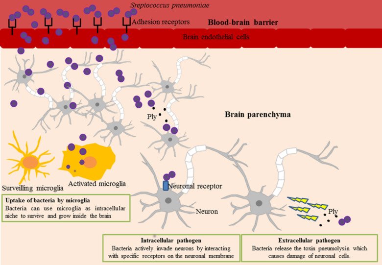
Pneumococcal infections of the brain.
Photo: Federico Iovino
After trafficking across the blood-brain barrier endothelium, S. pneumoniae in the brain encounters neurons and microglia. Neuronal damage can be caused by a direct interaction with the bacteria as a consequence of bacterial adhesion and invasion of neuronal cells, but also by an indirect interaction through the release of the toxin pneumolysin; microglia, the resident immune cells of the brain, have the fundamental function of eliminating bacteria by phagocytosis, on the other hand bacteria can also use microglia as niche to survive in the brain.
Projects
Neurons are the main cell component of the brain, and mediate many functions controlled by the brain. Neurological sequelae caused by bacterial infection of the brain occur in up to 70% of meningitis survivors and are due to a neuronal cell damage caused by the infection. Our goal is to study how pneumococci can interact with neurons, invade and kill neuronal cells.
- Scientists on this project: Davide Rizzato, Kristine Farmen
- Alumni on this project: Mahebali Tabusi, Hannah Haller
Publication
Neuronal death in pneumococcal meningitis is triggered by pneumolysin and RrgA interactions with β-actin.
Tabusi M, Thorsdottir S, Lysandrou M, Narciso AR, Minoia M, Srambickal CV, Widengren J, Henriques-Normark B, Iovino F
PLoS Pathog 2021 Mar;17(3):e1009432
Following up our recent discovery that ß-actin mediates pneumococcal binding to neuronal plasma membrane, we are now investigating novel approaches to block the interaction of S. pneumoniae with neuronal ß-actin to protect neurons during bacterial meningitis pathogenesis.
- Scientist on this project: Miguel Tofiño Vian
Neurons, as well as other eukaryotic cells, use autophagy to degrade accumulated and misfolded proteins and other waste material. Brain endothelial cells were previously shown to eliminate intracellular pneumococci through lysosomes. We want to investigate how neurons can use autophagy to eliminate intracellular pneumococci and protect themselves from bacterial infections during meningitis pathogenesis.
- Scientist on this project: Kristine Farmen, Lisa Knörr
Publication
Streptococcus pneumoniae invades endothelial host cells via multiple pathways and is killed in a lysosome dependent manner.
Gradstedt H, Iovino F, Bijlsma JJ
PLoS One 2013 ;8(6):e65626
Pneumolysin is a pore-forming toxin released by Streptococcus pneumoniae and is known to damage the eukaryotic cells that gets in contact with. Pneumolysin is a conserved protein among pneumococcal serotypes, however it is not known if several isoforms of pneumolysin are released by different strains of S. pneumoniae. We want to investigate if different meningitis clinical isolates express and release different pneumolysins, and if different isoforms of pneumolysin have different pore-forming activity which can lead to different degrees of cytotoxicity towards eukaryotic cells, neurons in particular.
- Scientist on this project: Simona Serra
- Alumni on this project: Vittorio Iannotti
Publication
The single D380 amino acid substitution increases pneumolysin cytotoxicity toward neuronal cells
Serra S, Iannotti V, Ferrante M, Tofiño-Vian M, Baxendale J, Silberberg G, Kohler TP, Hammerschmidt S, Ulijasz AT, Iovino F
iScience 27, 109583; April 19, 2024, https://doi.org/10.1016/j.isci.2024.109583
Microglia, the resident macrophages of the brain, are the primary line of defense for the brain towards infection. Microglia can recognize bacterial proteins either secreted or surface-exposed and initiate phagocytosis to actively eliminate pathogens. In this project, we want to identify the pneumococcal proteins that trigger microglial phagocytic activity and use them as immunostimulatory agents to enhance this bacterial clearing activity.
- Scientists on this project: Kristine Farmen, Johanna Hollenbeck
Publication
The Role of Microglia in Bacterial Meningitis: Inflammatory Response, Experimental Models and New Neuroprotective Therapeutic Strategies.
Thorsdottir S, Henriques-Normark B, Iovino F
Front Microbiol 2019 ;10():576
Using our established bacteremia-derived meningitis mouse model and whole brain microscopy imaging, we want to identify what are the brain regions that are mostly affected by blood-borne pneumococcal invasion upon penetration of the blood-brain barrier.
- Scientisits on this project: Kristine Farmen, Miguel Tofiño Vian
Publication
Spatio-temporal brain invasion pattern of Streptococcus pneumoniae and dynamic changes in the cellular environment in bacteremia-derived meningitis.
Farmen K, Tofiño-Vian M, Wellfelt K, Olson L, Iovino F
Neurobiol Dis 2024 Apr;195():106484
Many molecular processes, like the injury of neuronal cells and the neuro-inflammation process, are common in both bacterial meningitis and neurodegenerative diseases. We want to investigate to what extent the neuronal damage caused by a bacterial infection of the brain can increase the risk for onset of dementia, such as Alzheimer's disease.
- Scientisits on this project: Kristine Farmen, Miguel Tofiño Vian
Publication
Neuronal Damage and Neuroinflammation, a Bridge Between Bacterial Meningitis and Neurodegenerative Diseases.
Farmen K, Tofiño-Vian M, Iovino F
Front Cell Neurosci 2021 ;15():680858
Bacteriophage-encoded endolysins represent a novel type of antimicrobials that have received increasing attention due to their high killing efficacy, high specificity for the target pathogen and low chance of resistance development. Endolysins are enzymes encoded by bacteriophages that are produced at the end of the phage’s lytic cycle inside the bacterial host cell, from which they gain access to the cell wall and hydrolytically degrade their peptidoglycan substrate. In the context of Streptococcus pneumoniae, the antimicrobial potential has been demonstrated both in vitro and in vivo for many pneumococcal endolysins, such as the well-studied cpl-1, cpl-7 and PAL. However, a major knowledge gap remains regarding their application potential for pneumococcal meningitis. The aim of the research group is to preclinically characterize and evaluate these promising pneumococcal endolysins against clinically relevant pneumococcal meningitis clinical isolates using cell culture models in vitro and our established bacteremia-derived meningitis mouse model.
- Scientist on this project: Niels Vander Elst
The glymphatic system is responsible for solute exchange and clearance is a critical process to maintain proper brain homeostasis and take away from the brain harmful material. During pneumococcal meningitis, the glymphatic system's functionality is impaired and the lack of proper solute exchange process leads to a progressive accumulation in the brain of bacteria and bacterial toxic components with consequent neuroinflammation and neuronal damage. In this project, we investigate how physiological processes of the brain are altered during cerebral invasive infectious diseases and how we can translate our results into new therapeutic strategies to preserve solute exchange systems to protect the brain during infections.
- Alumni on this project: Sigrun Thórsdóttir
Publication
Dysfunctional Glymphatic System with Disrupted Aquaporin 4 Expression Pattern on Astrocytes Causes Bacterial Product Accumulation in the CSF during Pneumococcal Meningitis.
Generoso JS, Thorsdottir S, Collodel A, Dominguini D, Santo RRE, Petronilho F, Barichello T, Iovino F
mBio 2022 Oct;13(5):e0188622
Staff and contact
Group leader
- Federico IovinoPrincipal Researcher | Docent
All members of the group
- Simona SerraPhd Student
- Niels Vander ElstPostdoctoral Researcher
- Georgia YfantiResearch Assistant
Visiting address
Karolinska Institutet, Neuroscience, Solnavägen 9, Stockholm, 17165, Sweden
Student interns

Clara Caceffo

Valentina Mocci

Salem Tesfay
Contact us
Contact information for the Iovino Laboratory at the Department of Neuroscience, Karolinska Institutet.
Postal address
Karolinska Institutet
Department of Neuroscience
SE-171 77 Stockholm
Delivery address (goods, parcels, etc.)
Tomtebodavägen 16
SE-171 65 Solna