Our research
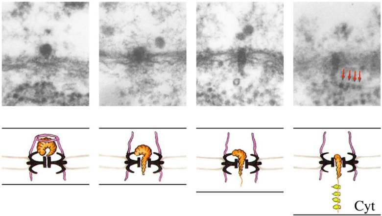
For many years we have studied how the genetic information is transferred from the genes in the cell nucleus to the polysomes in the cytoplasm in the context of the intact cell. In our experimental system, the salivary glands of the dipteran Chironomus tentans, it has been possible in a unique manner to visualise in the electron microscope the assembly and transport of a specific messenger ribonucleoprotein (mRNP) particle. We are now focusing on the passage of the mRNP particle through the nuclear pores and use an advanced electron microscopy method to study the molecular objects in 3D (electron tomography).
The mRNP particle has been isolated and characterised in 3D on the level of the elementary RNP fibril. This analysis forms the basis for the present in situ analysis of the initial interaction between the particle and the filaments of the nuclear basket. We have recently obtained data suggesting that the basket filaments seem to repel rather than attract particles; only those binding firmly to the nuclear pore complex remain bound resulting in an increased efficiency of transport of export-competent complexes. Our objective is now to use electron tomography to determine 1) whether the mRNP particles bind specifically to the basket filaments or to the core of the NPC, and 2) what part of the mRNPs interact with the binding sites.

Translocation of a messenger RNP particle through the nuclear pore. The particle binds to the basket, docks in front of the central channel, unfolds and passes through the central channel and finally exits into the cytoplasm and engages in translation.
We also take part in electron tomographic analysis of synaptonemal complexes in meiotic cells (with Abrahan Hernandez and Christer Höög) and desmosomes in human skin (with Lianne den Hollander and Lars Norlén) and in the development of the electron tomography method (with Ozan Öktem).
