Neural differentiation as a strategy for neuroblastoma treatment – Marie Arsenian Henriksson Group

We conduct research on Neuroblastoma, the most common extra cranial solid tumour of childhood that results in the highest number of cancer-related deaths in infants, as well as Medullobalstoma tumours.

Aula Medica_fotograf_Erik Flyg

Neural differentiation as a strategy for neuroblastoma treatment

Neuroblastoma is the most common extra cranial solid tumor of childhood that results in the highest number of cancer-related deaths in infants. In the high-risk group, approximately 40 percent of the patients are incurable despite intense multimodal treatment regiments. Amplification of the MYCN oncogene is strongly associated to poor survival and to an undifferentiated phenotype.

Considerable research efforts have been made to explore agents that could induce differentiation as therapeutic options for the high-risk patient group. So far, only retinoic acid treatment has shown promising results and today 13-cis retinoic acid is used as maintenance therapy.

MYCN belongs to the MYC network of transcription factors that plays a key role in the regulation of cell growth, apoptosis and differentiation. Other family members include c-MYC and L-MYC. All three genes are activated in a wide variety of human tumors.

While the c-MYC gene is expressed in most tissues, MYCN expression is restricted to early stages of embryonic development, making it a strong candidate as a potential therapeutic target. Importantly, inhibition of MYCN in neuroblastoma cells leads to differentiation, suggesting an important role for MYCN in maintaining an undifferentiated phenotype.

We have recently demonstrated that the MYCN-regulated miR-17~92 cluster targets several other nuclear hormone receptors (NHRs) in addition to ERalpha (Ribeiro et al, 2016). The glucocorticoid receptor (GR) emerged as particularly interesting. We found that it is a direct target of the miR-17~92 cluster, that it is the most significantly downregulated NHR in MYCN amplified neuroblastoma patients and is highly prognostic for patient outcome. Glucocorticoids, the hormones that bind to GR and promote its activation, have been successfully used to treat many diseases due to their anti-inflammatory, anti-proliferative, pro-apoptotic and anti-angiogenic properties. We found that low GR expression in was associated with an undifferentiated phenotype and decreased patient survival. Importantly, we showed that MYCN inhibition and subsequent reactivation of GR signaling promotes neural differentiation and reduces tumor burden. Our findings reveal a key role for the miR-17~92-regulated NHRs in neuroblastoma biology, thereby providing a potential differentiation approach for treating neuroblastoma patients (Ribeiro et al, 2016).

Together, our research will generate new insights into the pathology and the regulation of neural differentiation of MYCN-amplified neuroblastoma cells. This knowledge may offer novel prediction and diagnostic markers and serve as basis for development of new cancer therapies for children with neuroblastoma and other tumors since MYC is activated in many different cancer types.

News from Marie Arsenian Henriksson Group

Support our research

Publications

Selected publications

Funding

We are indebted to the following organizations for their valuable support for our research! Thank you!

Funding

Funds from the Karolinska Institutet

Staff and contact

Group leader

All members of the group

Former Group Members 

  • PostDoc Elena Eyre Sánchez
  • Associated Researcher Jenny Wilson
  • PostDoc Johanna Dzieran
  • PostDoc Nils Eickhoff
  • PostDoc Aine Henley
  • Associated Naomi Nagy
  • Researcher Ingibjorg Sigvaldadottir
  • PostDoc Diogo Ribeiro
  • Researcher Valentin Coronil
  • Associated Rashidul Islam
  • Researcher Anastasia Magoulopoulou
  • Undergraduate Student, Associated Malin Forss
  • PostDoc Theresa Wahlström
  • Affiliated researcher Sebastian Utz
  • Postdoc Wen Liu
  • Associated researcher Ulrica Westermark
  • Assistant Professor Anna Frenzel
  • PhD Jakob Lovén
  • Ami Albihn
  • Jiang Guosheng
  • Alexandre Drapier
  • Nikita Popov
  • Johan Ohlsson
  • Kenth Andersson
  • Jerome Marijsse
  • Conny Mathay
  • Ying Yang
  • Hao Mo
  • Marianne Crespin
  • Özgün Özer
  • Pinelopi Engskog Vlachos
  • Marcus Ladds
  • Somsundar Veppil Muralidharan
  • Sara Joanna Kritikos
  • Ina-Maria Rudolph
  • Shabana Zaidi
  • Madlen Jüttner
  • Research Student Rafael Galupa
  • Master student Marcus Klarqvist
  • Post Doc Karin Larsson
  • Research Associate Eric Fredlund
  • Biomedical Technician Roel Van Eijk

Michael Landreh Project

Principal Investigator Michael Landreh

Open Mass spectrometry of protein interactions from cancer to memory configuration options

Our research group focuses on the use of mass spectrometry (MS), a technique that allows us to determine the exact weight of biomolecules, to study how proteins recognise and bind their partners.

Mass spectrometry of protein interactions from cancer to memory

All biological processes can be described as biomolecules “talking” to each other, providing cargo, information, or transportation. These events usually take the from of a direct physical contact, i.e. a non-covalent interaction, in which one molecule, most often a protein, binds to one or more partners, inducing a change in the three-dimensional structure. In this manner, proteins can keep in touch with their environment to control their function. For example, upon sensing a change in pH, sider silk proteins lock each other into infinite chains to form very stable scaffolds, and membrane proteins can recognise individual lipid molecules in their environment to tune their activity accordingly. Aberrant, -faulty- interactions, on the other hand, interfere with these processes and are therefore often associated with diseases. Some proteins interact with themselves and form toxic structures such as amyloid fibrils that eventually lead to degeneration of the affected tissue, as seen in e.g Alzheimer's disease. Similarly, destabilization and aggregation of the tumour suppressor p53 and its targets leads loss of cell cycle control and impaired DNA damage repair, giving rise to cancer. Therefore, it is important to understand how exactly proteins “talk” to each other, and use this information to find ways to prevent interactions from going wrong.

Our group focuses on the use of mass spectrometry (MS), a technique that allows us to determine the exact weight of biomolecules, to study how proteins recognise and bind their partners. MS is well-suited for the study of transient interactions, large complexes and even unstable proteins, all of which are refractory to other structural biology methods like NMR and X-ray crystallography. For this purpose, we combine several complementary approaches:

  • In “native” MS, we gently transfer proteins together with their binding partners from physiological solutions into the vacuum inside the mass spectrometer and measure the weight and stability of the resulting complex. This reveals what type of interaction holds the partners together, and how many (and which) molecules are involved.
  • Hydrogen/deuterium exchange MS measures the incorporation of a chemical label (Deuterium) into the protein. Deuterium is incorporated into flexible and exposed parts of the protein. By measuring the resulting increase in weight, we are able to determine the stability and folding state of a protein, and even locate binding sites for tother proteins.
  • MS-based proteomics allows us to identify individual proteins from complex mixtures based on their unique mass “fingerprints”. Using individual proteins as bait, we are able to fish out their specific interaction partners and map upstream and downstream targets.

The combination of all three techniques provides direct insights into several aspects of an interaction, but also generates constraints that can be used to direct computational modelling.

Group Projects

Aggregation-prone proteins are related to a number of diseases such as neurodegenerative disorders, hereditary cancers, and interstitial lung disease, yet remain among the most challenging targets for structural biology. Many of these proteins use liquid-liquid phase separation (LLPS) to form membraneless organelles, but how the assembly of partially disordered proteins generates functional compartments in the cytoplasm and particularly in the nucleus is poorly understood.

Our group uses native mass spectrometry and ion mobility to probe the structures of proteins in membraneless organelles by releasing them from their native assemblies inside the mass spectrometer. Mass spectrometry data are complemented with atomistic and coarse-grained MD simulations, cryo-electron microscopy, and AI-guided structure predictions, to yield detailed models of proteins in native assemblies. We aim to assemble complete models of membraneless organelles that can open new avenues for drug development in cancer and neurodegeneration.

Membrane proteins constitute up to 60% of all drug targets, yet their location in the cell membrane makes studies of their native structures and interactions difficult. We use MS to understand how lipids affect the stability and function of proteins in the lipid bilayer. Current projects include the use of designed membrane proteins to extract first principles of lipid recognition, and how specific lipids can tune the internal dynamics of their target. Together with David Drew and Erik Lindahl at Stockholm University, we investigate the roles of lipids in transporters and ligand-gated ion channels.

A significant part of this work is the development of new mass spectrometric methods, as well as fundamental studies on the mechanisms of ionization and structures of proteins in the gas phase. We routinely use protein engineering and computational chemistry to find new ways to study protein structures and interactions by mass spectrometry.

We collaborate with David Drew and Erik Lindahl (Stockholm U) on membrane protein-lipid interactions, Erik Marklund (Uppsala) on integrating MS and MD simulations, Carol Robinson and Justin Benesch (Oxford) on MS method development, and Jan Johansson (KI) and Anna Rising (SLU) on strategies against amyloid formation, and the biology of spider silk production.

Publications

Neurodegenerative diseases and protein aggregation disorders

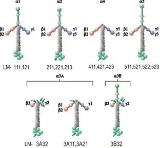
A strategy for the identification of protein architectures directly from ion mobility mass spectrometry data reveals stabilizing subunit interactions in light harvesting complexes.
Kaldmäe M, Sahin C, Saluri M, Marklund EG, Landreh M
Protein Sci. 2019 06;28(6):1024-1030

The microRNA miR-34 modulates ageing and neurodegeneration in Drosophila.
Liu N, Landreh M, Cao K, Abe M, Hendriks GJ, Kennerdell JR, et al
Nature 2012 Feb;482(7386):519-23

High-resolution structure of a BRICHOS domain and its implications for anti-amyloid chaperone activity on lung surfactant protein C.
Willander H, Askarieh G, Landreh M, Westermark P, Nordling K, Keränen H, et al
Proc. Natl. Acad. Sci. U.S.A. 2012 Feb;109(7):2325-9

The formation, function and regulation of amyloids: insights from structural biology.
Landreh M, Sawaya MR, Hipp MS, Eisenberg DS, Wüthrich K, Hartl FU
J. Intern. Med. 2016 Aug;280(2):164-76

Membrane proteins

Combining native and 'omics' mass spectrometry to identify endogenous ligands bound to membrane proteins.
Gault J, Liko I, Landreh M, Shutin D, Bolla JR, Jefferies D, Agasid M, Yen HY, Ladds MJGW, Lane DP, Khalid S, Mullen C, Remes PM, Huguet R, McAlister G, Goodwin M, Viner R, Syka JEP, Robinson CV
Nat Methods 2020 05;17(5):505-508

A Mass-Spectrometry-Based Approach to Distinguish Annular and Specific Lipid Binding to Membrane Proteins.
Bolla JR, Corey RA, Sahin C, Gault J, Hummer A, Hopper JTS, Lane DP, Drew D, Allison TM, Stansfeld PJ, Robinson CV, Landreh M
Angew Chem Int Ed Engl 2020 02;59(9):3523-3528

An engineered thermal-shift screen reveals specific lipid preferences of eukaryotic and prokaryotic membrane proteins.
Nji E, Chatzikyriakidou Y, Landreh M, Drew D
Nat Commun 2018 10;9(1):4253

Lipids Shape the Electron Acceptor-Binding Site of the Peripheral Membrane Protein Dihydroorotate Dehydrogenase.
Costeira-Paulo J, Gault J, Popova G, Ladds MJGW, van Leeuwen IMM, Sarr M, et al
Cell Chem Biol 2018 03;25(3):309-317.e4

The role of interfacial lipids in stabilizing membrane protein oligomers.
Gupta K, Donlan JAC, Hopper JTS, Uzdavinys P, Landreh M, Struwe WB, et al
Nature 2017 01;541(7637):421-424

Integrating mass spectrometry with MD simulations reveals the role of lipids in Na+/H+ antiporters.
Landreh M, Marklund EG, Uzdavinys P, Degiacomi MT, Coincon M, Gault J, et al
Nat Commun 2017 01;8():13993

Spider silk

A pH-dependent dimer lock in spider silk protein.
Landreh M, Askarieh G, Nordling K, Hedhammar M, Rising A, Casals C, et al
J. Mol. Biol. 2010 Nov;404(2):328-36

Biomimetic spinning of artificial spider silk from a chimeric minispidroin.
Andersson M, Jia Q, Abella A, Lee XY, Landreh M, Purhonen P, et al
Nat. Chem. Biol. 2017 03;13(3):262-264

Mass spectrometry captures structural intermediates in protein fiber self-assembly.
Landreh M, Andersson M, Marklund EG, Jia Q, Meng Q, Johansson J, et al
Chem. Commun. (Camb.) 2017 Mar;53(23):3319-3322

Mass spectrometric method development

Charge Engineering Reveals the Roles of Ionizable Side Chains in Electrospray Ionization Mass Spectrometry.
Abramsson ML, Sahin C, Hopper JTS, Branca RMM, Danielsson J, Xu M, Chandler SA, Österlund N, Ilag LL, Leppert A, Costeira-Paulo J, Lang L, Teilum K, Laganowsky A, Benesch JLP, Oliveberg M, Robinson CV, Marklund EG, Allison TM, Winther JR, Landreh M
JACS Au 2021 Dec;1(12):2385-2393

Mass Spectrometry Reveals the Direct Action of a Chemical Chaperone.
Gault J, Lianoudaki D, Kaldmäe M, Kronqvist N, Rising A, Johansson J, et al
J Phys Chem Lett 2018 Jul;9(14):4082-4086

A membrane cell for on-line hydrogen/deuterium exchange to study protein folding and protein-protein interactions by mass spectrometry.
Astorga-Wells J, Landreh M, Johansson J, Bergman T, Jörnvall H
Mol. Cell Proteomics 2011 Sep;10(9):M110.006510

Controlling release, unfolding and dissociation of membrane protein complexes in the gas phase through collisional cooling.
Landreh M, Liko I, Uzdavinys P, Coincon M, Hopper JT, Drew D, et al
Chem. Commun. (Camb.) 2015 Nov;51(85):15582-4

Low Charge and Reduced Mobility of Membrane Protein Complexes Has Implications for Calibration of Collision Cross Section Measurements.
Allison TM, Landreh M, Benesch JLP, Robinson CV
Anal. Chem. 2016 06;88(11):5879-5884

List of all of Michael Landreh's publications on PubMed


Contact

Profile image

Michael Landreh

Principal Researcher

David Lane Group

The protein p53, widely known as the guardian of the genome, was discovered by Prof. Sir David Lane in 1979 and has over the years been recognized as a tumor suppressor gene mutated in more than half of all malignant tumors occurring in adults. Apart from its role as a tumor suppressor, p53 has been shown to be involved in numerous regulatory cell functions. Research in our lab is focused on understanding the many facets of p53 biology in malignant and normal cells. We employ cutting edge technology such as mass spectometry, advanced microscopy, multi-color flow cytometry and imaging cytometry to this end and strive to translate our findings to the clinic.

Projects within the David Lane Group

Principal Investigator Michael Landreh

Publications

How to lose tumor suppression
David Philip Lane
Science 365 (6453), 539-540 2019 Aug;()

Understanding p53 functions through p53 antibodies.
Sabapathy K, Lane DP
J Mol Cell Biol 2019 Apr;11(4):317-329

The MDM2/MDMX-p53 Antagonist PM2 Radiosensitizes Wild-Type p53 Tumors.
Spiegelberg D, Mortensen AC, Lundsten S, Brown CJ, Lane DP, Nestor M
Cancer Res. 2018 Sep;78(17):5084-5093

Constitutive activation of WASp in X-linked neutropenia renders neutrophils hyperactive.
Keszei M, Record J, Kritikou JS, Wurzer H, Geyer C, Thiemann M, et al
J. Clin. Invest. 2018 Aug;128(9):4115-4131

Mass Spectrometry Reveals the Direct Action of a Chemical Chaperone.
Gault J, Lianoudaki D, Kaldmäe M, Kronqvist N, Rising A, Johansson J, et al
J Phys Chem Lett 2018 Jul;9(14):4082-4086

Monoclonal Antibodies against Specific p53 Hotspot Mutants as Potential Tools for Precision Medicine.
Hwang LA, Phang BH, Liew OW, Iqbal J, Koh XH, Koh XY, et al
Cell Rep 2018 01;22(1):299-312

Inhibiting p53 Acetylation Reduces Cancer Chemotoxicity.
Zheng S, Koh XY, Goh HC, Rahmat SAB, Hwang LA, Lane DP
Cancer Res. 2017 08;77(16):4342-4354

Exploiting the p53 Pathway for Therapy.
Cheok CF, Lane DP
Cold Spring Harb Perspect Med 2017 Mar;7(3):

Cancer. p53, guardian of the genome.
Lane DP
Nature 1992 Jul;358(6381):15-6

T antigen is bound to a host protein in SV40-transformed cells.
Lane DP, Crawford LV
Nature 1979 Mar;278(5701):261-3

Further publications

All scientific publications from David Lane

Funding

The Swedish Research Council (Vetenskapsrådet)

Contact

David Lane Laboratory at Sci Life
Biomedicum quarter 7B-C Solnavägen 9, 17165 Solna

P53 Laboratory in Singapore
The p53 lab in singapore is focusing on the development of new therapies, new diagnostics and new discoveries in the p53 pathway.
More information on the website

Manuel Patarroyo Project

The present research program investigates the role of laminins and their cell-surface receptors in tumor cell adhesion, migration, survival, self-renewal and proliferation as well as in tumor invasion, metastasis and chemoresistance.

The Laminin family
Figure 1. The Laminin family.

Laminins and their cell-surface receptors in tumor invasion and metastasis

Tumor invasion and metastasis accounts for most cancer-related deaths but their molecular basis is poorly understood. During the metastatic cascade tumor cells migrate, survive, self-renew and proliferate via interaction with extracellular matrix proteins such as laminins. Notably, this cell-matrix interaction also contributes to chemotherapy resistance.

Laminins, a large family of αβγ heterotrimeric proteins primarily found in basement membranes (Fig. 1), are masters of normal tissue architecture, a property which is highly disrupted during tumor invasion and metastasis. In addition to their structural functions, laminins promote cell adhesion, migration, survival, self-renewal and proliferation. Over 15 laminin isoforms are presently known and their expression is developmentally regulated and cell- and tissue-specific. Laminins, via their α chain, are differentially bound by integrins and other cell-surface receptors. Although expression of laminin isoforms in tumors mostly reflects expression in their normal counterparts, distinct alterations of laminin expression and function occur during tumor invasion, particularly in epithelial-mesenchymal transition of the tumors cells and loss of the basement membrane barrier. During local dissemination and metastasis cancer cells encounter exogenous laminins in blood/lymphatic vessels, nerves, lymphoid tissue and other anatomical structures. Moreover, the tumor cells themselves are able to produce and secrete laminins and to use these endogenous molecules in an autocrine fashion.

The present research program investigates the role of laminins and their cell-surface receptors in tumor cell adhesion, migration, survival, self-renewal and proliferation as well as in tumor invasion, metastasis and chemoresistance. Both tumor-derived (endogenous) and non-tumor (exogenous) laminins are studied as well as their effect on tumor cells at different stages, including cancer stem cells and following epithelial-mesenchymal transition. Examination of laminin isoforms in tumor tissues and biological fluids has a diagnostic/prognostic potential, and antagonists of laminin-receptor interactions may constitute novel therapeutic strategies against malignant diseases.

In parallel studies the role of laminin isoforms in immune/inflammatory responses and tissue repair is also investigated.

Publications

Laminin alpha 5 regulates mammary gland remodeling through luminal cell differentiation and Wnt4-mediated epithelial crosstalk.
Englund JI, Ritchie A, Blaas L, Cojoc H, Pentinmikko N, Döhla J, Iqbal S, Patarroyo M, Katajisto P
Development 2021 06;148(12):

Maternal alloimmune IgG causes anti-glomerular basement membrane disease in perinatal transgenic mice that express human laminin α5.
Abrahamson DR, Steenhard BM, Stroganova L, Zelenchuk A, St John PL, Petroff MG, et al
Kidney Int. 2019 12;96(6):1320-1331

HAX-1 overexpression in multiple myeloma is associated with poor survival.
Feng X, Kwiecinska A, Rossmann E, Bottai M, Ishikawa T, Patarroyo M, et al
Br. J. Haematol. 2019 04;185(1):179-183

Culturing functional pancreatic islets on α5-laminins and curative transplantation to diabetic mice.
Sigmundsson K, Ojala JRM, Öhman MK, Österholm AM, Moreno-Moral A, Domogatskaya A, et al
Matrix Biol. 2018 09;70():5-19

Tetraspanin CD151 and integrin α6β1 mediate platelet-enhanced endothelial colony forming cell angiogenesis.
Huang Z, Miao X, Patarroyo M, Nilsson GP, Pernow J, Li N
J. Thromb. Haemost. 2016 Mar;14(3):606-18

Mesenchymal Stromal Cells for Sphincter Regeneration: Role of Laminin Isoforms upon Myogenic Differentiation.
Seeger T, Hart M, Patarroyo M, Rolauffs B, Aicher WK, Klein G
PLoS ONE 2015 ;10(9):e0137419

Bacterial genotoxins promote inside-out integrin β1 activation, formation of focal adhesion complexes and cell spreading.
Levi L, Toyooka T, Patarroyo M, Frisan T
PLoS ONE 2015 ;10(4):e0124119

A laminin 511 matrix is regulated by TAZ and functions as the ligand for the α6Bβ1 integrin to sustain breast cancer stem cells.
Chang C, Goel HL, Gao H, Pursell B, Shultz LD, Greiner DL, et al
Genes Dev. 2015 Jan;29(1):1-6

Laminins 411 and 421 differentially promote tumor cell migration via α6β1 integrin and MCAM (CD146).
Ishikawa T, Wondimu Z, Oikawa Y, Gentilcore G, Kiessling R, Egyhazi Brage S, et al
Matrix Biol. 2014 Sep;38():69-83

Monoclonal antibodies to human laminin α4 chain globular domain inhibit tumor cell adhesion and migration on laminins 411 and 421, and binding of α6β1 integrin and MCAM to α4-laminins.
Ishikawa T, Wondimu Z, Oikawa Y, Ingerpuu S, Virtanen I, Patarroyo M
Matrix Biol. 2014 Jun;36():5-14

Platelets store laminins 411/421 and 511/521 in compartments distinct from α- or dense granules and secrete these proteins via microvesicles.
Pook M, Tamming L, Padari K, Tiido T, Maimets T, Patarroyo M, et al
J. Thromb. Haemost. 2014 Apr;12(4):519-27

A novel monoclonal antibody to human laminin α5 chain strongly inhibits integrin-mediated cell adhesion and migration on laminins 511 and 521.
Wondimu Z, Omrani S, Ishikawa T, Javed F, Oikawa Y, Virtanen I, et al
PLoS ONE 2013 ;8(1):e53648

Integrin-mediated adhesion of human mesenchymal stem cells to extracellular matrix proteins adsorbed to polymer surfaces.
Dånmark S, Finne-Wistrand A, Albertsson AC, Patarroyo M, Mustafa K
Biomed Mater 2012 Jun;7(3):035011

Melanoma cells produce multiple laminin isoforms and strongly migrate on α5 laminin(s) via several integrin receptors.
Oikawa Y, Hansson J, Sasaki T, Rousselle P, Domogatskaya A, Rodin S, et al
Exp. Cell Res. 2011 May;317(8):1119-33

Project Members

The project currently has vacant research positions, if you are interested please contact Manuel Patarroyo on the email address below

Contact