Wilhelm lab

Our aim is to understand mechanisms regulating cancer and how the cross talk between tumor cells, the surrounding stroma and infiltrating immune cells control tumor development. We use reprogramming techniques to generate disease-relevant stem cell models that mimic the disease to uncover mechanisms that turn healthy cells into cancer cells and to uncover novel targets for developing future therapies.

Group members

Mechanisms regulating childhood tumors medulloblastoma and neuroblastoma

Medulloblastoma and Neuroblastoma are among the most common malignant neural tumors in children. Neural tumors constitute around one third of all childhood cancers, but almost half of the mortalities. Although advances in therapies have increased survival of the patients, many of the survivors experience complications due to the harsh treatment. This shows not only a need for increasing our understanding of molecular mechanisms operating during neural tumor formation, but furthermore, it highlights the importance of developing targeted therapies that will spare the developing child while specifically eradicating tumor cells.

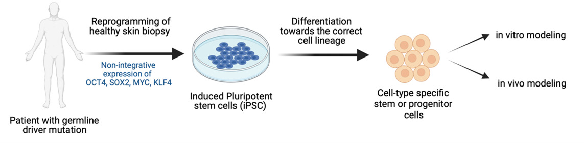
To achieve this, we have developed new cancer models using human disease-relevant cell types. By somatic cell reprogramming to induced pluripotent stem (iPS) cells and differentiation to neural stem cells, we are developing new cancer models with cells from patients with familial driver mutations known to cause medulloblastoma and neuroblastoma.

Wilhelm lab, graphic illustrations of reprogramming of cells.
Reprogramming of cells. Photo: BioRender.com
Wilhelm lab, graphic illustrations of models for identifying molecular mechanisms.
Models for identifying molecular mechanisms. Photo: BioRender.com

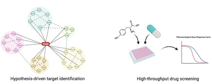
Identification of novel therapeutic targets

We use our models for identifying molecular mechanisms regulating the disease but also for finding targets that could be used for precision cancer medicine. We combine hypothesis-driven approaches based on mechanistic studies with unbiased exploratory compound library screens to identify novel targets. Targets are validated using our cellular models, in vivo models and in patient material.

Wilhelm lab, graphic illustration of cerebellar organoid derived from iPS cells
Cerebellar organoid derived from iPS cells. PAX6 (red), Tuj1 (green). Photo: Niek van Bree

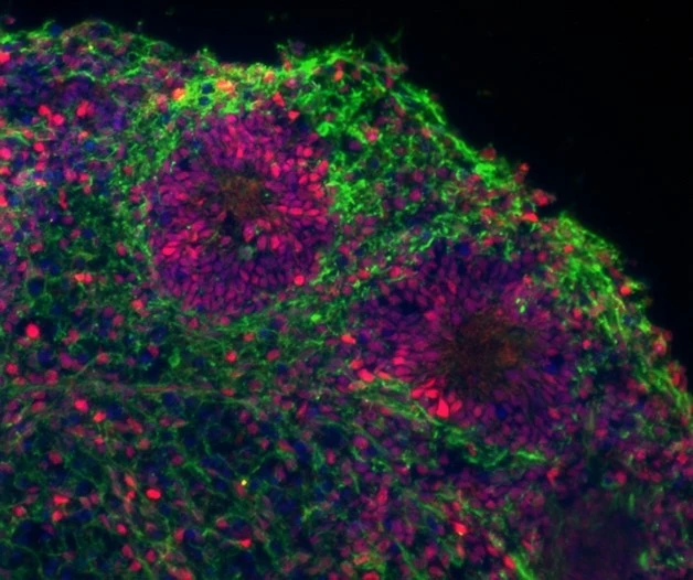
Develop ex-vivo neural organoid models

To create more sophisticated ex-vivo models, we are generating human brain organoids by differentiating iPS cells towards the neural lineage allowing for 3D self-organisation into brain-like structures. This allows us to study how cancer driver-mutations initiate tumor development, how transformed cells interact with neighboring cells, and provides a better model system to evaluate drug responses.

Wilhelm lab, graphic illustrations
Patrolling gd T cells engaged in killing tumor cells. Photo: Lola Boutin

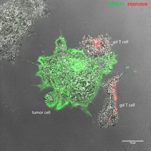
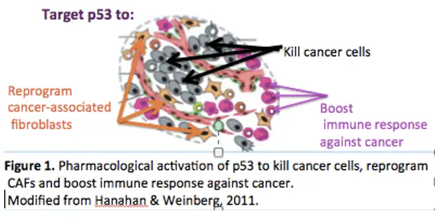
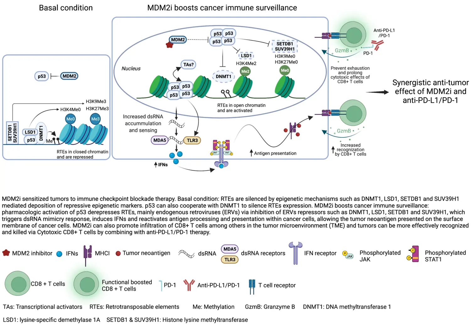
The role of microenvironment in tumor development and how it can be used for cancer therapy

Our lab has a long-standing interest in the cross-talk between tumors cells and the microenvironment, and we have previously studied how tumor angiogenesis, hypoxic responses, drug resistance mechanisms, and immune responses are regulated.

Currently, we are interested how the age of the surrounding environment may affect tumor development. In addition, we are investigating the potential of using non-HLA restricted T cells, also called non-conventional T lymphocytes (NC-T cells), for adoptive cell therapy to eliminate tumors while sparing healthy tissue.

Vacancies

The Wilhelm lab are always looking for motivated Postdocs, please apply using contact information below.

Open positions for Doctoral students are posted on KI homepage when available.

Support our research

Publications

Selected publications

Staff and contact

Group leader

All members of the group

Contact and visit us

Contact information for the Wilhelm lab at the Department of Microbiology, Tumor and Cell Biology, Karolinska Institutet.

Postal address

Karolinska Institutet
Department of Microbiology, Tumor and Cell Biology
171 77 Stockholm

Visiting address (visitors, couriers, etc.)

Karolinska Institutet
Biomedicum, B7
Solnavägen 9
171 65 Solna

Delivery address (goods, parcels, etc.)

Tomtebodavägen 16
171 65 Solna

Where to find us

News

Alumni

PhD students

  • Johanna Wolfsberger, PhD, Defended her thesis 2020-11-05
  • Ana Marin Navarro, PhD, Defended her thesis 2019-03-08
  • Habib Sakil, PhD, Defended his thesis 2017-10-05

Postdocs

  • Veronica Zubillaga, PhD, Postdoc 2019-2020
  • Marina Stantic, PhD, Postdoc 2012-2018, Assistant Professor 2018-2019
  • Evelyn Susanto, PhD, Postdoc 2013-2018

Project students 

  • Maria-Luisa Wiesinger, Master student, DKFZ Heidelberg University, 2021-2022
  • Maria Calvo Noriega, Master student, Karolinska Institutet, 2021-2022
  • Nicola Bell, Bachelor student, Hochschule Zitau/Görlitz, 2021
  • Paloma Araceli Ruiz de Castroviejo Teba, Master student, Karolinska Institutet, 2021
  • Elena Baldisseri, Project student, Karolinska Institutet, 2020
  • Silvia Schäfer, Master student, Albert-Ludwigs-Universität Freiburg, Germany, 2020
  • Akhilesh Kapdi, Bachelor student, University of Applied Science Krems, 2019
  • Mark Tan Kia, Bachelor student, King's College London, 2019
  • Lennart Sänger, Master student, Martin Luther University Halle , 2018-2019
  • Okan Gültekin, Master student, Karolinska Institutet, 2018
  • Aitor Bermejo, Project student, Karolinska Institutet, 2018
  • Astrid van der Geest, Master student, Utrecht University, 2018
  • Stefanie Renken, Master student, Karolinska Institutet, 2017
  • Larsen Vornholz, Mater student, Karolinska Institutet, 2017
  • Janina Henze, Master student, Frankfurt University, 2017
  • Jonne Rietdjik, Master student, Radboud University, 2017
  • Marleen Meyer, Master student, 2015
  • Haizea Goni, Master student, Karolinska Institutet, 2015
  • Nikolina Giotopoulou, Master student, Karolinska Institutet, 2014
  • Jannis Kalkitsas, Master student, Karolinska Institutet, 2014
  • Trixy Fang, Master student, Karolinska Institutet, 2014
  • Atra Barsham, Master student, Karolinska Institutet, 2013
  • Elin Edsbäcker, Master student, Karolinska Institutet, 2013
  • Rafael Galupa, Master student, Karolinska Institutet, 2012
  • Anja Waldman, Master student, Lübeck University, 2012
  • Martina Czapko, Master student, 2011
  • Rebecca Khan, Bachelor student, Karolinska Institutet, 2011

Kaisa Lehti Group

Tumor Cell – Microenvironment Communication in Cancer Invasion, Metastasis and Drug Resistance

Cancer metastasis and recurrence rely on tumor dissemination and tissue (re)colonization by interchangeable modes of cell invasion and growth. Such cellular plasticity also contributes to anticancer drug resistance, representing a major challenge in cancer research and clinical practice. Multiple tumor microenvironment (TME) communication pathways have been discovered. However, it remains incompletely understood how tumor cells integrate changes in cell-cell interactions, soluble factors and cues or physical confines of the extracellular matrix (ECM) with cytoskeletal dynamics and intracellular signaling to produce TME-dependent, plastic responses controlling growth, invasion and drug resistance.

To date, our results have revealed distinct mechanisms of TME-dependent oncogenic receptor tyrosine kinase signalling, ECM remodelling and transmembrane protease - adhesion receptor crosstalk.

Our Projects

I. The fibrotic and inflammatory responses in cancer

The extracellular matrix (ECM) is a tissue compartment of the tumor microenvironment (TME) that not only serves as a physical scaffold, but also influences cancer and stromal cells through biochemical and biophysical cues. During cancer progression and chemotherapy treatment, the TME undergoes major changes affecting ECM composition and biomechanical properties, inducing fibrosis and inflammation.

In our group, we have a long-term interest in membrane-type matrix metalloproteinases (MT-MMPs) that are involved in modulation of the TME, thereby regulating cancer cell proliferation, invasion and dissemination. Our previous studies have for instance have described a phenotypic switch from collagen infiltration to lymphovascular invasion, linear collagen assembly and tumor cell aggregation upon co-expression of MT1-MMP/MMP14 and MT3-MMP/MMP16 in the most aggressive melanoma tumors.

In our current study, we investigate how the matrisome (a group of genes encoding core ECM proteins and ECM-associated proteins, including cytokines, chemokines and growth factors) changes upon disease progression and chemotherapy treatment in patients with ovarian high grade serous carcinoma (HGSC). In addition, we explore how fibrotic matrisome changes upon HGSC metastasis and chemotherapy can alter cancer cell functions and vice versa how the cancer cell alterations affect the ECM sensing and remodeling.

Relevant Publications

Fibroblasts in the Tumor Microenvironment: Shield or Spear?
Alkasalias T, Moyano-Galceran L, Arsenian-Henriksson M, Lehti K
Int J Mol Sci 2018 May;19(5):

Membrane-type matrix metalloproteases as diverse effectors of cancer progression.
Turunen SP, Tatti-Bugaeva O, Lehti K
Biochim Biophys Acta Mol Cell Res 2017 Nov;1864(11 Pt A):1974-1988

MMP16 Mediates a Proteolytic Switch to Promote Cell-Cell Adhesion, Collagen Alignment, and Lymphatic Invasion in Melanoma.
Tatti O, Gucciardo E, Pekkonen P, Holopainen T, Louhimo R, Repo P, et al
Cancer Res 2015 May;75(10):2083-94

Actin-associated protein palladin promotes tumor cell invasion by linking extracellular matrix degradation to cell cytoskeleton.
von Nandelstadh P, Gucciardo E, Lohi J, Li R, Sugiyama N, Carpen O, et al
Mol Biol Cell 2014 Sep;25(17):2556-70

II. Receptor tyrosine kinase signaling in cancer

The transmission of extracellular signals into the cell and to the nucleus ensuring downstream cellular responses, i.e. signaling transduction, allows cell-cell communication within varying TMEs. Receptor tyrosine kinases (RTK) are membrane receptors that sense extracellular cues and promote diverse signaling responses regulating normal cellular processes. In cancer, these receptors are frequently altered to promote malignant cancer cell behavior and thereby explored as targets for current and future therapies.

Using a genome-wide gain-of-function human kinome screen we identified novel MT1-MMP  regulators, including two RTKs, namely fibroblast growth factor receptor 4 (FGFR4) and Eph receptor A2 (EphA2). Our studies have shown that cancer-associated FGFR4 polymorphism affects the activity of an FGFR4-MT1-MMP complex in cancer progression, and that an EphA2-MT1-MMP axis regulates cancer invasion. In addition, we have uncovered a mechanism of FGFR4 oncogenic activity via suppression of the stress-associated mammalian sterile20-like kinases (MST1/2)-induced apoptosis machinery in tumor cells with prominent HER/ERBB and FGFR4 signaling-driven proliferation.

Most recently we identified a switch in EphA2 signaling induced upon treatment of ovarian cancer cells with platinum chemotherapy and linked to increased treatment resistance. By inhibiting the kinases mediating this oncogenic signaling (RSK1/2), we could efficiently sensitize cells to the chemotherapy-induced apoptosis in 2D and 3D models of HGSC ex vivo as well as in vivo, presenting a novel approach of how to improve currently available therapies.

Based on these results, our ongoing research explores the mechanisms of the complex signaling crosstalk governed by FGFRs and the context-dependent Eph-ephrin system, as well as the regulation of these signals by interactions with both the MMPs and adhesion molecules in human ovarian breast and gastric carcinomas.

Relevant Publications

Adaptive RSK-EphA2-GPRC5A signaling switch triggers chemotherapy resistance in ovarian cancer.
Moyano-Galceran L, Pietilä EA, Turunen SP, Corvigno S, Hjerpe E, Bulanova D, et al
EMBO Mol Med 2020 04;12(4):e11177

Crosstalk Between Cancer Associated Fibroblasts and Cancer Cells in Scirrhous Type Gastric Cancer.
Miki Y, Yashiro M, Moyano-Galceran L, Sugimoto A, Ohira M, Lehti K
Front Oncol 2020 ;10():568557

FGFR4 phosphorylates MST1 to confer breast cancer cells resistance to MST1/2-dependent apoptosis
Turunen SP, Von Nandelstadh P, Öhman T, Gucciardo E, Seashore-ludlow B, Martins B, Rantanen V, Li H, Höpfner K, Östling P, Varjosalo M, Lehti K Cell death and differentiation 2019;26(12):2577-2593

Eph- and ephrin-dependent mechanisms in tumor and stem cell dynamics
Gucciardo E, Sugiyama N, Lehti K Cellular and molecular life sciences: CMLS 2014;71(19):3685-710

EphA2 cleavage by MT1-MMP triggers single cancer cell invasion via homotypic cell repulsion
Sugiyama N, Gucciardo E, Tatti O, Varjosalo M, Hyytiäinen M, Gstaiger M, Lehti K The Journal of cell biology 2013;201(3):467-84

EphA2 bears plasticity to tumor invasion.
Sugiyama N, Gucciardo E, Lehti K
Cell Cycle 2013 Sep;12(18):2927-8

FGF receptor-4 (FGFR4) polymorphism acts as an activity switch of a membrane type 1 matrix metalloproteinase-FGFR4 complex.
Sugiyama N, Varjosalo M, Meller P, Lohi J, Chan KM, Zhou Z, et al
Proc Natl Acad Sci U S A 2010 Sep;107(36):15786-91

Fibroblast growth factor receptor 4 regulates tumor invasion by coupling fibroblast growth factor signaling to extracellular matrix degradation.
Sugiyama N, Varjosalo M, Meller P, Lohi J, Hyytiäinen M, Kilpinen S, et al
Cancer Res 2010 Oct;70(20):7851-61

III. IPLA-OVCA

Project leader Sahar Salehi

In the Lehti lab, we use modern molecular and cell biology techniques to examine how surgical treatment, the extent of surgery and local anesthetics affect mesothelial inflammation and tumour cell phenotype and functions in epithelial ovarian cancer (EOC). Moreover, the effects of identified molecular pathways on aggressive tumour properties such as invasion and chemotherapy resistance of EOC cells are tested in functional ex vivo assays.

By prospectively collecting relevant tumour and blood samples from our clinical Phase III trial (IPLA-OVCA) (https://clinicaltrials.gov/ct2/show/NCT04065009), translational investigation of the key molecules and cellular mechanisms underlying the unfavorable patient responses to the surgical trauma are investigated in detail with the specific aims:

I. To examine if surgical peritoneal stress evolves during the course of surgery, as measured by pro-inflammatory cytokines and biomarkers in the mesothelium, tumour tissues and blood, coupled to analysis of the circulating tumour DNA

II. To test if intraperitoneally administered anti-inflammatory analgesics may reduce the (mesothelial) inflammation.

III. To use functional ex vivo assays for exploring how the above identified surgical tissue responses and induced or inhibited pathways affect the aggressive EOC cell functions and properties including tumour invasion and chemo-resistance. 

The samples are transported fresh and directly from the operating theatre Karolinska University Hospital to the Lehti lab, KI. Each included patient generates close to 300 subsamples after processing.

Epithelial ovarian cancer (EOC) constitute cancers with origin in the fallopian tube, ovary or peritoneum and is the gynecologic malignancy with the highest mortality rate. Most women are diagnosed with advanced stage disease (70%) at presentation, when EOC has exfoliated to the peritoneal cavity. In Sweden, 650 women are diagnosed annually. Strategies for prolonging lives of women with EOC, are widening with emerging targeted therapies. Albeit this development, surgery remains the cornerstone in the available treatment armamentarium. Undisputedly, radical and complete surgical resection of tumour in combination with medical treatment (platinum-based chemotherapy) confers the basis for best survival outcomes in patients with EOC. For this reason, an abrupt shift in surgical treatment algorithm to aggressive upfront radical surgery with high proficiency surgeons was implemented Karolinska University Hospital (KUH) in 2014. The Stockholm Ovarian Cancer Project (STOOVCA) began as a quality assurance project in women with advanced EOC in the Stockholm/Gotland Region. The aim was to assess surgical quality and outcomes (survival) before and after this abrupt shift in surgical treatment algorithm to aggressive upfront ultra-radical surgery. Our hypothesis for this clinical study was that a shift to primary ultra-radical surgery in a population confers superior survival in patients subjected to surgery, without compromising the total overall survival regardless of primary treatment. Contrary to expectation we found that a shift to ultra-radical upfront surgery in a population did not improve overall survival in surgically treated patients, and instead conferred an inferior 3-year survival in patients subjected to radical surgery.

In our other recently completed clinical randomized double-blinded and placebo-controlled pilot study, we hypothesized that due to the known anti-inflammatory effect of local anesthetics (LA), postoperative recovery would be facilitated. In 40 patients undergoing cytoreductive surgery, randomized to treatment with either perioperative intraperitoneal local anesthetics (experiment) or saline (control), we found that the time-interval from surgery to start of adjuvant chemotherapy, an endpoint associated with prolonged survival, was significantly reduced in the intraperitoneal local anesthetics group.

For this reason, further investigation with a an adequately powered Phase III trial started recruitment in September 2020.

Relevant publications

Ultra-radical upfront surgery does not improve survival in women with advanced epithelial ovarian cancer; a natural experiment in a complete population.
Falconer H, Joneborg U, Krawiec K, Palsdottir K, Bottai M, Salehi S
Gynecol Oncol 2020 Oct;159(1):58-65

Intraperitoneal ropivacaine reduces time interval to initiation of chemotherapy after surgery for advanced ovarian cancer: randomised controlled double-blind pilot study.
Hayden JM, Oras J, Block L, Thörn SE, Palmqvist C, Salehi S, et al
Br J Anaesth 2020 05;124(5):562-570

IV. Sarcoma tumor microenvironment 

Project leader Jordi Gonzalez-Molina

Sarcomas are a very diverse group of malignancies accounting for about 1% of adult and 15% of childhood cancers. Sarcomas arise from connective tissues and are typically rich in structural extracellular matrix (ECM) proteins such as fibrillar collagens. With the final goal of improving patient treatment, we are investigating the impact of the tumour microenvironment on sarcomagenesis and tumour development. In close collaboration with Dr Joseph Carlson’s team, we have recently identified the ECM and the tumour immune microenvironment (TIME) as potentially interregulated factors with prognostic value in uterine sarcomas. This has motivated us to further investigate the function of the ECM in sarcoma. We are currently working in two main sarcoma projects:

I. Impact of collagen biomechanics and microarchitecture on rhabdomyosarcoma metastasis potential.

II. Evolution and impact of the ECM and the TIME on uterine mesenchymal tumour development.

These studies involve an in-depth tissue characterisation combining multi-omics technologies with biomechanical and image analysis techniques. Moreover, we develop biomaterial-based 3D culture systems to mimic clinically relevant tissue features to functionally assess the mechanisms of action of potential novel biomarkers and therapeutic targets.

Picture from Kaisa Lehti group
Picture from Kaisa Lehti group.

Relevant Publications

The extracellular fluid macromolecular composition differentially affects cell-substrate adhesion and cell morphology.
Gonzalez-Molina J, Mendonça da Silva J, Fuller B, Selden C
Sci Rep 2019 06;9(1):8505

Integrated Molecular Analysis of Undifferentiated Uterine Sarcomas Reveals Clinically Relevant Molecular Subtypes.
Binzer-Panchal A, Hardell E, Viklund B, Ghaderi M, Bosse T, Nucci MR, et al
Clin Cancer Res 2019 04;25(7):2155-2165

Extracellular fluid viscosity enhances liver cancer cell mechanosensing and migration.
Gonzalez-Molina J, Zhang X, Borghesan M, Mendonça da Silva J, Awan M, Fuller B, et al
Biomaterials 2018 09;177():113-124

MMP14 in Sarcoma: A Regulator of Tumor Microenvironment Communication in Connective Tissues.
Gonzalez-Molina J, Gramolelli S, Liao Z, Carlson JW, Ojala PM, Lehti K
Cells 2019 08;8(9):

Contact

Profile image

Kaisa Lehti

Researcher

Galina Selivanova Group

p53 reactivation illustration
Photo: n/a

Manipulation of the p53 tumor suppressor pathway: from lab bench to clinic

p53 is the major tumor suppressor which eliminates damaged or oncogene-expressing cells by activating transcription of genes inducing apoptosis, cell cycle arrest or senescence. p53 inactivation via mutations or enhanced degradation by MDM2 is the most frequent alteration in human cancers, which underscores the key role of p53 in combating cancer. Reinstatement of p53 by genetic means have demonstrated remarkable tumor suppression in animal models, including inhibition of aggressive metastatic lesions. This inspires the idea of developing small molecules reactivating p53 to fight cancer (Figure 1).

Illustration Figure 2. strategy of p53 restoration by small molecules depends on the type of p53 inactivation
Figure 2. strategy of p53 restoration by small molecules depends on the type of p53 inactivation. Photo: n/a

Depending on the type of p53 inactivation, there could be envisioned two major strategies of p53 reinstatement: restoring the function of mutant p53 and preventing p53 inactivation by MDM2 (Figure 2).

A number of MDM2 inhibitors reactivating wild type p53 have been discovered, including small molecule RITA discovered by us (Issaeva et al, Nature Medicine, 2004; Enge et al, Cancer Cell 2009; Grinkevich et al, Cancer Cell, 2009).  Several of these are currently being tested in clinical trials, including derivatives of nutlin and the stapled peptide ATSP-7041, which are activating wild type (wt) p53 are in Phase I/II trials (www.cilicaltrials.gov, see also our review Sanz G et al, J Mol Cell Biol, 2019).

Mutant p53 is expressed in cancers, where it adopts an unfolded conformation resulting in loss of DNA binding and in some cases gaining oncogenic function. Thus, our hypothesis is to stabilize p53 conformation by small molecule to restore the DNA binding tumor suppressor function of p53. Since around 50% of all human tumors carry mutations in p53, it could be widely applicable in clinic. We have identified a small molecule PRIMA-1MET by screening NCI chemical library using cell-based assay (Bykov et al, Nature Medicine, 2002). PRIMA-1MET(commercial name APR-246, please see www.aprea.com) restores the active conformation and DNA binding of mutant p53 in cells and in vitro, re-activates the function of mutant p53 in tumor cells of different origin and suppreses the growth of human xenograft tumors in mice.  

APR-246 has been tested in patients in Phase I clinical trial and in March 2020 APR-246 received Fast Track and Breakthrough Therapy designations from FDA based on outstanding results of Phase II trial in MDS patients. These prompted the start of Phase III randomized trial in MDS.

p53 targets are working on different levels: to prevent tumor development by inducing growth arrest and DNA repair as well as anioxidant response or to exterminate developed tumors by inducing senescence, apoptosis and immunologic cell death. Photo: Galina Selivanova och Alexandra Selivanova

p53 targets are working on different levels: to prevent tumor development by inducing growth arrest and DNA repair as well as anioxidant response or to exterminate developed tumors by inducing senescence, apoptosis and immunologic cell death. Photo: Galina Selivanova och Alexandra Selivanova

In light of clinical developments of p53-targeting therapies, it is imperative to rationally design combinatorial treatments (since monotherapy cannot cure cancer) and find biomarkers to stratify patients. To achieve these, we need to get a deep understanding of the molecular mechanisms and pathways affected by p53 and p53-reactivating compounds.

Illustration
Figure 1. Pharmacological activation of p53 to kill cancer cells, reprogram CAFs and boost immune response against cancer. Photo: Galina Selinova

Projects

Targeting p53 to kill tumor cells, reprogram cancer-associated fibroblasts and boost anti-cancer immune response

Half of human tumors carry mutations in the p53 gene, resulting in the expression of inactive protein. Tumors that do not carry p53 mutations, develop an alternative mechanisms of p53 inactivation, converging on enhanced proteasomal degradation. Given the extraordinary high frequency of p53 inactivation in tumors and the high potency of p53 in elimination of tumors, it appears highly desirable to restore the tumor suppressor function of p53 as a strategy to combat cancer. A number of p53-targeting therapies are currently being tested in patients. 

We are addressing the fundamental questions that need to be solved for the efficient application of p53-targeting medicines and their combinations in clinic, i.e. thorough understanding of the mechanism of action of the identified compounds, including target specificity in vitro and in vivo and off-target effects.

While p53 reactivating molecules have been shown to kill cancer cells, the question remains open how p53 reinstatement will affect tumor microenvironment, cancer-associated fibroblasts in particular, as well as anti-cancer immune response. 

Our ambition is to provide basis for innovative p53-based treatment strategies targeting both cancer cells and tumor microenvironment to generate synergistic anti-tumor effects that result in long term survival of patients. Defining this concept, its mechanisms and implications for novel anti-cancer therapies form the main objectives of our studies.

In addition to applied aspects, we are actively working on important basic aspects of tumor biology, including genetic screens for p53 regulators and identification of novel factors which control p53 activity in tumor and in normal cells, which can serve as targets for therapeutic intervention in a future. In essence, to understand p53 is to understand how its interaction with proteins, and thus DNA, is controlled. In order to address these challenging questions we will employ a comprehensive and multidisciplinary approach. We will apply cutting-edge molecular and cell biology methodologies, multi-omics analyses and systems biology analysis, combined with ex vivo 3D cell & tissue culture and in vivo models. We combine hypothesis-driven strategy with unbiased multi-omics approach and apply the analysis of publicly available patient data sets (e.g., TCGA), as well as newly obtained data from patient material via my collaborations with clinicians in Sweden and abroad.

We perform highly parallel and comprehensive search for p53 modulators and identify their functional significance by inter-disciplinary integration of genome-wide expression profiling, ChIP-seq and proteomic approaches, followed by systems biology analysis. This will pave the way to the identification of key p53 target genes and factors contributing to alternative biological responses. These will be thoroughly validated in cells, mouse models and patient samples using functional genomics and protein-protein interaction assays. Selected factors will be used for chemical libraries screens to identify small molecules that target them.  We hope our studies will open the way for the development of novel therapeutic approaches.

In a recent study published in Cancer Discovery in 2021, we demonstrated that pharmacological activation of p53 induces the expression of ERVs and generation of double-stranded (ds) RNA which caused intracellular dsRNA stress leading to type 1 and type 3 IFN responses and induction of  antigen processing and presentation  (APP) genes.  Notably, we found that p53 activation promotes the recruitment of immune cells to tumors in vivo in mouse models and sensitizes refractory tumors to programmed cell death protein 1 (PD-1) blockade.  

Figure illustrating MDM2 inhibitors sensitize tumors to Immune checkpoint blockade.
MDM2 inhibitors sensitize tumors to Immune checkpoint blockade. Photo: Xiaolei Zhou

Importantly, the analysis of pre- and post-treatment tumor biopsy samples from melanoma patients treated with the experimental MDM2 inhibitor ALRN-6924,  revealed the induction of viral mimicry response genes, as well as immune function signatures suggesting infiltration of cytotoxic CD8+ T cells.

Our results suggest the potential role of  p53-reactivating compounds with checkpoint inhibitors. In conclusion, our data presented across cancer cell lines, tumor-bearing mouse models, and melanoma patients suggest that pharmacological p53 reactivation triggers the ERV-dsRNA-IFN pathway within tumor cells, thereby altering the tumor microenvironment evoking tumor immune surveillance.

Selected publications

Conference publication: CANCER SCIENCE. 2023;114:249
Pharmacological activation of p53 triggers viral mimicry response thereby promoting anti-tumor immunity Selivanova G; Zhou X; Singh M; Sanz G; Guerlavais V; Carvajal LA; Aivado M; Annis A; Zhan Y; Oliveira M; Westerberg L; Johnsen JI

Article: BRITISH JOURNAL OF CANCER. 2022;127(11):2060-2071
Mutant p53 gain of function mediates cancer immune escape that is counteracted by APR-246
Zhou X; Santos GS; Zhan Y; Oliveira MMS; Rezaei S; Singh M; Peuget S; Westerberg LS; Johnsen JI; Selivanova G

Article: MOLECULAR CANCER THERAPEUTICS. 2022;21(10):1524-1534
Decreased DNA Damage and Improved p53 Specificity of RITA Analogs
Zhan Y; Zhou X; Peuget S; Singh M; Peyser BD; Fan Z; Selivanova G

Article: ONCOGENE. 2022;41(15):2173-2186
Enterobacteria impair host p53 tumor suppressor activity through mRNA destabilization
Aschtgen M-S; Fragkoulis K; Sanz G; Normark S; Selivanova G; Henriques-Normark B; Peuget S

Article: FRONTIERS IN MOLECULAR BIOSCIENCES. 2022;9:823195
Novel Allosteric Mechanism of Dual p53/MDM2 and p53/MDM4 Inhibition by a Small Molecule Grinkevich VV; Vema A; Fawkner K; Issaeva N; Andreotti V; Dickinson ER; Hedstrom E; Spinnler C; Inga A; Larsson L-G; Karlen A; Wilhelm M; Barran PE; Okorokov AL; Selivanova G; Zawacka-Pankau JEAll authors

Editorial comment: CANCERS. 2021;13(19):4850
p53-Dependent Repression: DREAM or Reality? Peuget S; Selivanova G

Article: ISCIENCE. 2020;23(12):101785
Class IV Lasso Peptides Synergistically Induce Proliferation of Cancer Cells and Sensitize Them to Doxorubicin Guerrero-Garzon JF; Madland E; Zehl M; Singh M; Rezaei S; Aachmann FL; Courtade G; Urban E; Rueckert C; Busche T; Kalinowski J; Cao Y-R; Jiang Y; Jiang C-L; Selivanova G; Zotchev SBAll authors

Article: BREAST CANCER RESEARCH. 2020;22(1):80
Identification and targeting of selective vulnerability rendered by tamoxifen resistance
Singh M; Zhou X; Chen X; Santos GS; Peuget S; Cheng Q; Rihani A; Arner ESJ; Hartman J; Selivanova G

Article: CANCER RESEARCH. 2020;80(7):1538-1550 
Thermal Proteome Profiling Identifies Oxidative-Dependent Inhibition of the Transcription of Major Oncogenes as a New Therapeutic Mechanism for Select Anticancer Compounds Peuget S; Zhu J; Sanz G; Singh M; Gaetani M; Chen X; Shi Y; Saei AA; Visnes T; Lindstrom MS; Rihani A; Moyano-Galceran L; Carlson JW; Hjerpe E; Joneborg U; Lehti K; Hartman J; Helleday T; Zubarev R; Selivanova GAll authors

Article: JOURNAL OF CLINICAL MEDICINE. 2020;9(2):E598-598
p53 CRISPR Deletion Affects DNA Structure and Nuclear Architecture Rangel-Pozzo A; Booth S; Yu PLI; Singh M; Selivanova G; Mai S

Article: SCIENTIFIC REPORTS. 2020;10(1):1049
Pifithrin-α alters p53 post-translational modifications pattern and differentially inhibits p53 target genes Zhu J; Singh M; Selivanova G; Peuget S

Article: HAEMATOLOGICA. 2020;105(1):170-181
Therapeutic targeting of mutant p53 in pediatric acute lymphoblastic leukemia Demir S; Boldrin E; Sun Q; Hampp S; Tausch E; Eckert C; Ebinger M; Handgretinger R; te Kronnie G; Wiesmueller L; Stilgenbauer S; Selivanova G; Debatin K-M; Meyer LHAll authors

Article: CELL DEATH & DISEASE. 2019;10(11):845
RITA requires eIF2α-dependent modulation of mRNA translation for its anti-cancer activity
Ristau J; van Hoef V; Peuget S; Zhu J; Guan B-J; Liang S; Hatzoglou M; Topisirovic I; Selivanova G; Larsson O

Review: JOURNAL OF MOLECULAR CELL BIOLOGY. 2019;11(7):586-599
Inhibition of p53 inhibitors: progress, challenges and perspectives Sanz G; Singh M; Peuget S; Selivanova G

Contact

Profile image

Sylvain Peuget Group

p53 regulation by bacteria and its impact on tumorigenesis  

Increasing evidence from metagenomics study highlights the role of the microbiota composition in cancer initiation, progression and resistance to therapies. Cancers are clearly associated with bacterial dysbiosis, which could be a passenger but also a driver of the tumorigenesis process. However, little is known about how the bacteria species enriched during dysbiosis promote tumorigenesis.

We are investigating the molecular mechanisms of the host response to bacterial dysbiosis, with a focus on the tumor suppressor p53 pathway. p53 plays a central role in cell signaling and is therefore strongly regulated in response to commensal and pathogenic bacteria signaling during inflammation. Moreover, p53 encodes a transcriptional program which in turns regulates the innate and adaptive immune response. Therefore, it is not surprising that bacteria have evolved different mechanisms to target and manipulate the host p53 pathway in order to establish and keep their niche. Finally, p53 is the major barrier against cancer in humans and its role as a regulator of immune response is emerging as a key component for its tumor suppressive function. Hence, bacterial interference with the p53 pathway could have important consequences for tumorigenesis.

Main questions 

  1. How the p53 pathway is involved in the physiological host response to bacteria, i.e. inflammation and innate immune response?  
  2. How some bacteria hijack the p53 pathway for their own benefits?  
  3. How bacteria-induced deregulation of the p53 pathway influence cancer initiation and progression?  

Our ultimate goal is to find new therapeutic targets in bacteria and/or the human host to help prevent of combat cancer.  

To investigate these fundamental questions at the molecular level both on the bacteria and host side, this project is a collaboration performed together with an expert in microbiology of pathogenic bacteria Ass. Prof. Marie-Stephanie Aschtgen in Prof Birgitta Henriques-Normark’s lab.

p53 regulation by bacteria and its impact on tumorigenesis

Contact

Profile image

Sylvain Peuget

Research Specialist

Ingemar Ernberg Group

Our primary research interest is infection and cancer in humans.  We particularly study Epstein-Barr Virus (EBV)-infection, which can also be used as a model to study early steps in in vitro transformation, viral genetic switches, non-genetic variation and cancer cell metabolism.

Ingemar Ernberg Group

Contact

Profile image

Ingemar Ernberg

Professor, Senior