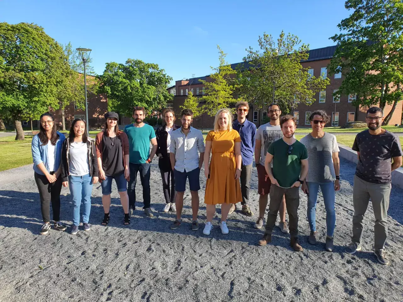
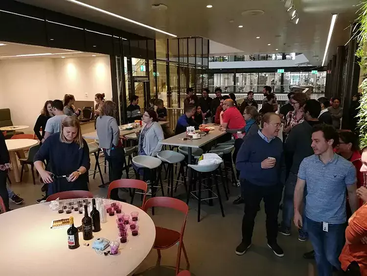
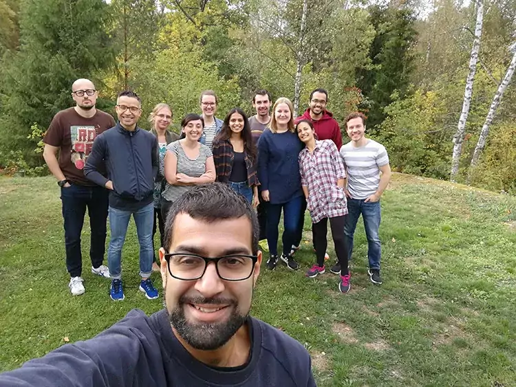
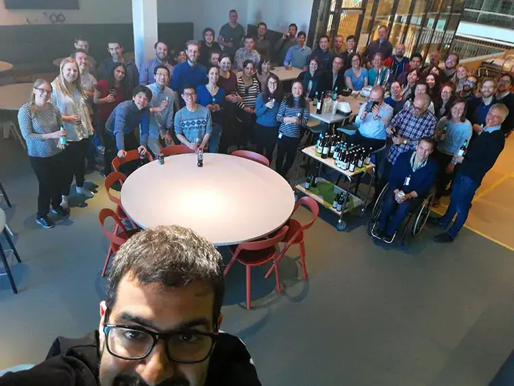
Gonçalo Castelo-Branco Group

Our research group is interested in the molecular mechanisms defining the epigenetic state of cells of the oligodendrocyte lineage. We are focused on how interplay between transcription factors, non-coding RNAs and chromatin modifying enzymes contribute to the transition between epigenetic states within the oligodendrocyte lineage, with the aim to design epigenetic based-therapies to induce regeneration (remyelination) in demyelinating diseases, such as multiple sclerosis.

Pictures from Goncalo Castelo Branco Group Lab retreat in Visby 2023

Our research

Our research group is interested in the molecular mechanisms defining the transcriptomic and epigenomic states of oligodendrocyte lineage cells. We are particularly focused on how interplay between transcription factors, non-coding RNAs and chromatin modifying enzymes contributes to the transition between epigenetic states in oligodendroglia, with the aim to design epigenetic based-therapies to induce regeneration/remyelination and prevent neuroinflammation in demyelinating diseases, such as MS.

Illustration of oligodendocyte progenitor cells.
Binary choices of oligodendrocyte progenitor cells (OPCs), differentiation/myelination or transition to an immune state. Photo: Amagoia Agirre

Research area

All cells in a given organism are derived from a single cell (zygote) and thereby share an identical genome. Additional layers of epigenetic information overlaid on the genome achieve the plethora of cellular phenotypes present in development and in the adult body. This epigenetic information is stored at the level of chromatin, the complex where nuclear DNA is packaged together with histones. DNA methylation and post-translational modifications at histones define the epigenetic state of a cell and ultimately cell fate, by controlling key processes, including transcription. Non-coding RNAs have also emerged recently as key regulators of chromatin and cell fate.

Oligodendrocytes insulate neuronal axons through their myelin containing membranes. Myelin allows the fast and efficient impulse transmission between neurons through saltatory conduction and is important for axonal integrity, thereby being essential for the proper functioning of the central nervous system.

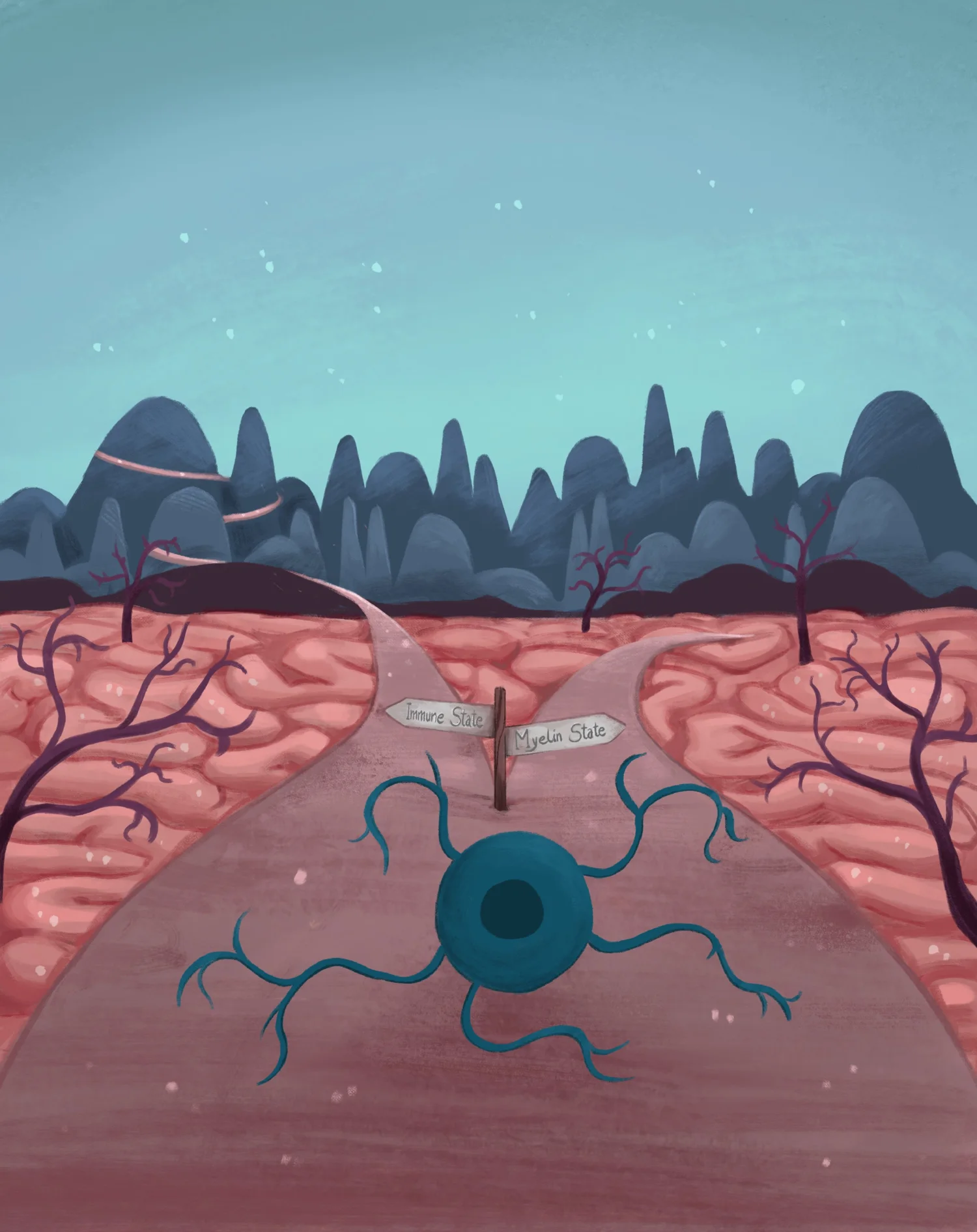
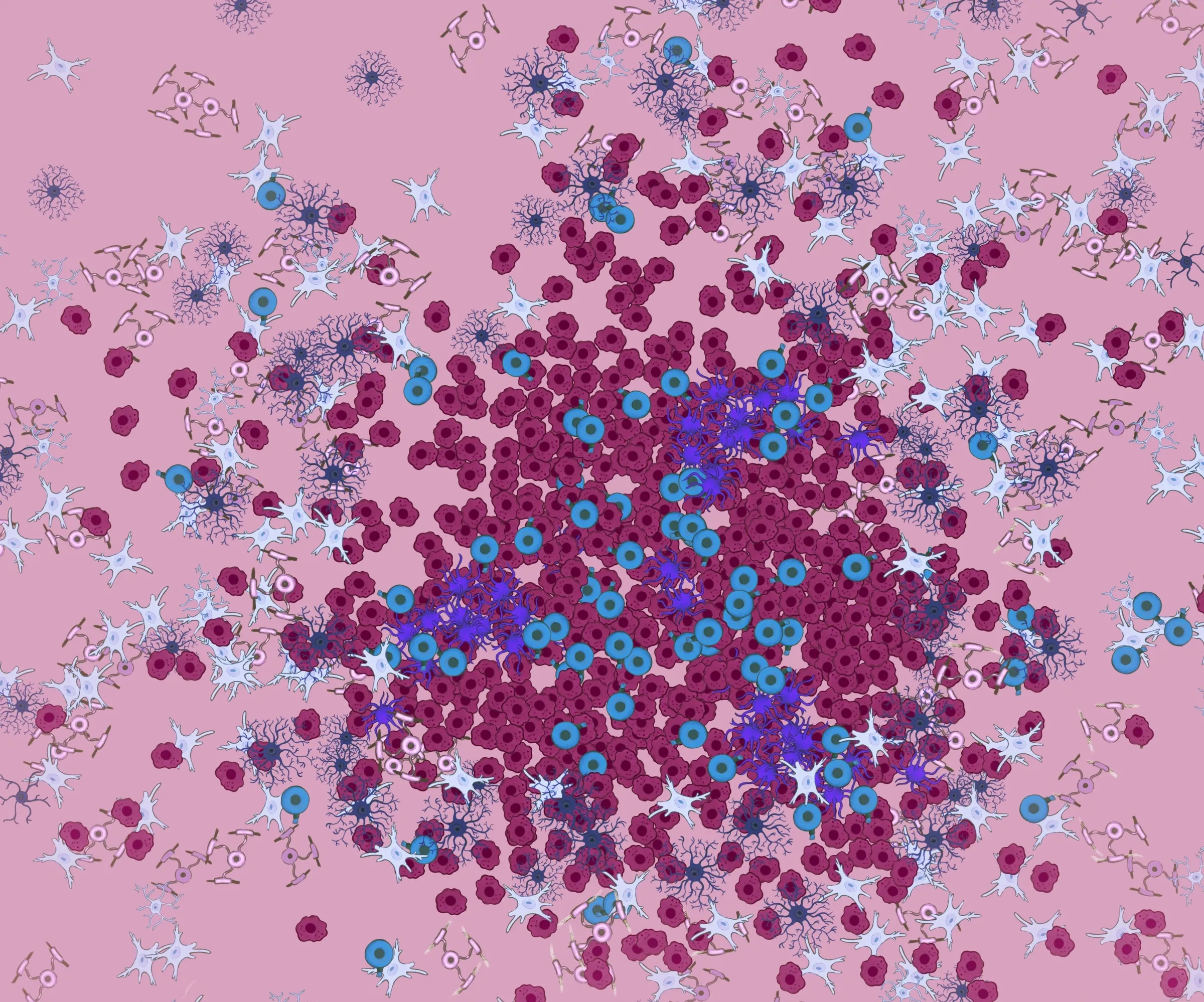
Several diseases, such as multiple sclerosis (MS), are characterized by abnormal or defective myelination. Spontaneous remyelination occurs at initial stages of MS, promoted by endogenous oligodendrocyte precursor cells (OPCs). However, this process progressively starts occurring with less efficiency, until it eventually fails. Oligodendrocyte precursors (OPCs) start to be specified early during embryogenesis, in different areas of the embryonic brain, but their terminal differentiation and functional maturation occurs only at post-natal stages. The epigenetic state of OPCs define their ability to remain as a precursor cell, differentiate or even de-differentiate into a stem cell state or a glioma initiating cell state.

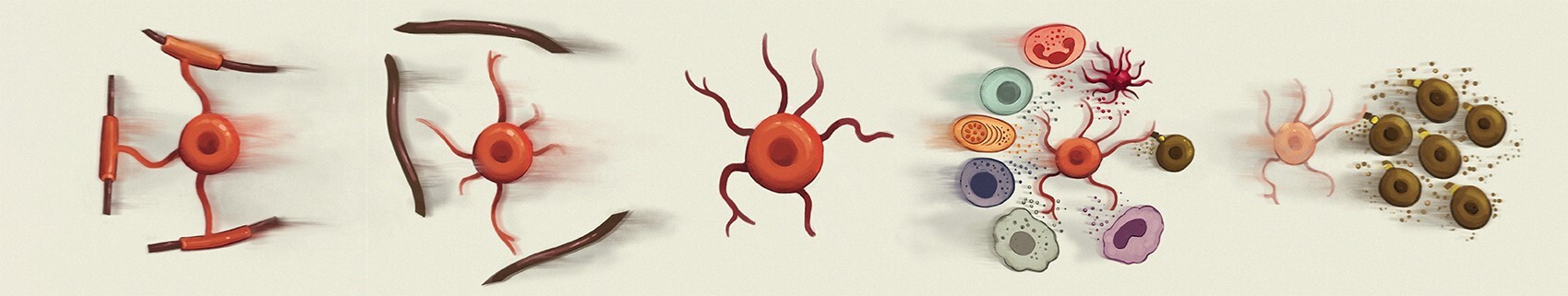
The main focus of our research group is to investigate how different epigenetic states in oligodendroglia  (OPCs and oligodendrocytes) are established, by identifying key chromatin modifying complexes and non-coding RNAs that are involved in epigenetic transitions, using technologies such as single-cell and spatial transcriptomics and epigenomics, among others. We identified novel distinct oligodendrocyte cell states during differentiation/myelination (Science 2015, Science 2016, Nature Communications 2020) and also during development (Developmental Cell, 2018, Developmental Cell 2022). We have also expanded our single-cell omics approach to the MOG EAE mouse model of MS and to human patient samples, in which we could discriminate distinct disease-associated OPC/OL states (Nature Medicine, 2018, Nature 2019, bioRxiv 2022).

We have implemented scATAC-Seq, which allows examination of chromatin accessibility at a single cell level, applying for instance to examine the oligodendrocyte lineage in the EAE mouse model of MS (Neuron 2022). We found that oligodendroglia is already primed at an epigenomic level at immune genes, to allow their transcription in inflammatory environments. We also found that some susceptibility variant for MS are located in regions of open chromatin in human and mouse oligodendroglia, which might implicates these cells in MS aetiology and progression.

We are very interested in technological development in single-cell and spatial epigenomics. We have developed single-cell CUT&Tag to examine the simultaneous profile of histone modifications at a single-cell level in large number of cells and applied to investigate the epigenomic heterogeneity of the mouse brain (Nature Biotechnology 2021). We also developed a multimodal and optimized iteration of scCUT&Tag called nano-CT (for nano-CUT&Tag) that allows simultaneous probing of three epigenomic modalities at single-cell resolution, using nanobody-Tn5 fusion proteins (Nature Biotechnology 2022). In collaboration with Prof. Rong Fan at Yale University, we have implemented CUT&Tag and ATAC-Seq at a spatial level, by using a new ligation-based method for deterministic barcoding in tissue, probing different histone modifications and chromatin accessibility in the mouse brain (Nature 2022 and Science 2022).

We generated several web resources from our single-cell and spatial omic datasets, compiled in our OligoInternode interface, where one can enter your gene of interest and investigate its expression pattern in the identified populations and cell states, including the oligodendrocyte lineage. One can explore how a gene of interest is differentially expressed or the chromatin landscape in regulatory regions surrounding your gene of interest.

Selected latest publications

Multimodal chromatin profiling using nanobody-based single-cell CUT&Tag.
Bartosovic M, Castelo-Branco G
Nat Biotechnol 2022 Dec;():

Spatial profiling of chromatin accessibility in mouse and human tissues.
Deng Y, Bartosovic M, Ma S, Zhang D, Kukanja P, Xiao Y, Su G, Liu Y, Qin X, Rosoklija GB, Dwork AJ, Mann JJ, Xu ML, Halene S, Craft JE, Leong KW, Boldrini M, Castelo-Branco G, Fan R
Nature 2022 Sep;609(7926):375-383

Spatial-CUT&Tag: Spatially resolved chromatin modification profiling at the cellular level.
Deng Y, Bartosovic M, Kukanja P, Zhang D, Liu Y, Su G, Enninful A, Bai Z, Castelo-Branco G, Fan R
Science 2022 Feb;375(6581):681-686

Epigenomic priming of immune genes implicates oligodendroglia in multiple sclerosis susceptibility
Mandy Meijer*, Eneritz Agirre*, Mukund Kabbe, Cassandra A. van Tuijn, Abeer Heskol, Chao Zheng, Ana Mendanha Falcão, Marek Bartosovic, Leslie Kirby, Daniela Calini, Michael R. Johnson, M. Ryan Corces, Thomas J. Montine, Xingqi Chen, Howard Y. Chang, Dheeraj Malhotra, and Gonçalo Castelo-Branco
Neuron 2022 doi: 10.1016/j.neuron.2021.12.034

Single-cell CUT&Tag profiles histone modifications and transcription factors in complex tissues.
Bartosovic M, Kabbe M, Castelo-Branco G
Nat Biotechnol 2021 Apr;():
Access the article with SharedIT

Distinct oligodendrocyte populations have spatial preference and different responses to spinal cord injury.
Floriddia EM, Lourenço T, Zhang S, van Bruggen D, Hilscher MM, Kukanja P, et al
Nat Commun 2020 11;11(1):5860

Crossing boundaries: Interplay between the immune system and oligodendrocyte lineage cells.
Kirby L, Castelo-Branco G
Semin Cell Dev Biol 2020 Nov;():

Altered human oligodendrocyte heterogeneity in multiple sclerosis
Sarah Jäkel, Eneritz Agirre, Ana Mendanha Falcão, David van Bruggen, Ka Wai Lee, Irene Knuesel, Dheeraj Malhotra, Charles ffrench-Constant, Anna Williams and Gonçalo Castelo-Branco.
Nature. 2019 Feb;566(7745):543-547.

Disease-specific oligodendrocyte lineage cells arise in multiple sclerosis.
Falcão AM, van Bruggen D, Marques S, Meijer M, Jäkel S, Agirre E, et al
Nat. Med. 2018 12;24(12):1837-1844
Full text

Transcriptional Convergence of Oligodendrocyte Lineage Progenitors during Development.
Marques S, van Bruggen D, Vanichkina DP, Floriddia EM, Munguba H, Väremo L, et al
Dev. Cell 2018 08;46(4):504-517.e7

Oligodendrocyte heterogeneity in the mouse juvenile and adult central nervous system.
Marques S, Zeisel A, Codeluppi S, van Bruggen D, Mendanha Falcão A, Xiao L, et al
Science 2016 Jun;352(6291):1326-1329
Article at KI News: Nerve-insulating cells more diverse than previously thought

Publications

Selected publications

Members and contact

Group leader

All members of the group

Other people connected to the group

  • van, Rixel Esmée
  • Gentili, Mattia
  • de, Boer Fleur

News

Upcoming events

OligoInternode: datasets and resources

Our group has generated web resources from our single-cell, bulk and spatial transcriptomic and epigenomic datasets, where one can enter your gene of interest and investigate its expression pattern in the identified populations and cell states, including the oligodendrocyte lineage. One can explore how a gene of interest is differentially expressed or the chromatin landscape in regulatory regions surrounding your gene of interest.

Spatial Transcriptomics and Epigenomics

Dataset: Single cell-resolution in situ sequencing of spinal cords of the EAE mouse model of MS and human MS spinal cord

These spatial datasets accompany the manuscript below, in collaboration with Prof. Mats Nilsson, Stockholm University. They feature single-cell spatial expression profiling using In situ sequencing characterising the spatio-temporal dynamics of major cellular subtypes in EAE and MS spinal cord archival tissue (in this case using the Xenium in situ sequencing platform from 10X Genomics).

Cellular architecture of evolving neuroinflammatory lesions and multiple sclerosis pathology 
Petra Kukanja*, Christoffer M. Langseth*, Leslie A. Rubio Rodríguez-Kirby, Eneritz Agirre, Chao Zheng, Amitha Raman, Chika Yokota, Christophe Avenel, Katarina Tiklová, André O. Guerreiro-Cacais, Tomas Olsson, Markus M. Hilscher, Mats Nilsson, Gonçalo Castelo-Branco
Cell, online March 20, 2024, Cell, doi: 10.1016/j.cell.2024.02.030

Dataset: Spatial epigenome–transcriptome of mouse embryo E13.5, mouse juvenile brain (P22) and human adult hippocampus

These spatial datasets accompanies the manuscript below, in collaboration with Prof. Rong Fan, Yale University, US. They feature dBit-Seq, profiling simultaneously RNA and chromatin accessibility (ATAC), or RNA and CUT&Tag (for H3K27me3 (repressive mark), H3K4me3 (active mark) of H3K27ac (active mark), in several complex tissues.

Spatially resolved epigenome-transcriptome co-profiling of mammalian tissues
Di Zhang, Yanxiang Deng, Petra Kukanja, Eneritz Agirre, Marek Bartosovic, Mingze Dong, Cong Ma, Sai Ma, Graham Su, Shuozhen Bao, Yang Liu, Yang Xiao, Gorazd B. Rosoklija, Andrew J. Dwork, J. John Mann, Kam W. Leong, Maura Boldrini, Liya Wang, Maximilian Haeussler, Benjamin J. Raphael, Yuval Kluger, Gonçalo Castelo-Branco*, Rong Fan*
Nature 616, 113–122 (2023).

Dataset: Spatial chromatin accessibility of mouse embryo E13.5, mouse juvenile brain and human adult hippocampus

All datasets via Atlasxomic spatial browser

These spatial datasets accompanies the manuscript below, in collaboration with Prof. Rong Fan at Yale University. We have implemented ATAC-Seq at a spatial level, probing chromatin accessibility at a spatial level in complex tissue.

Spatial profiling of chromatin accessibility in mouse and human tissues.
Deng Y, Bartosovic M, Ma S, Zhang D, Kukanja P, Xiao Y, Su G, Liu Y, Qin X, Rosoklija GB, Dwork AJ, Mann JJ, Xu ML, Halene S, Craft JE, Leong KW, Boldrini M, Castelo-Branco G, Fan R
Nature 2022 Sep;609(7926):375-383

Dataset: Spatially resolved chromatin modification profiling of mouse embryo E13.5 and mouse juvenile brain

All datasets via Atlasxomic spatial browser

These spatial datasets accompanies the manuscript below, in collaboration with Prof. Rong Fan at Yale University. We have implemented CUT&Tag (for H3K27me3 (repressive mark) and H3K4me3 (active mark)) at a spatial level, probing different histone modifications in complex tissues.

Spatial-CUT&Tag: Spatially resolved chromatin modification profiling at the cellular level.
Deng Y, Bartosovic M, Kukanja P, Zhang D, Liu Y, Su G, Enninful A, Bai Z, Castelo-Branco G, Fan R
Science 2022 Feb;375(6581):681-686

Single Cell Transcriptomics and Epigenomics

Simultaneous single-cell multiome ATAC/ RNA.seq of oligodendroglia from the experimental autoimmune encephalomyelitis (EAE) MS mouse model at different stages of the disease course.

We applied simultaneous single-cell multiome ATAC and RNA sequencing to oligodendroglia FACS-sorted from male and female EAE at three distinct time points:1) early stage, when neurological symptoms begin to arise; 2) peak, after symptoms rapidly increased; 3) late stages, when the disease is chronic and progression has stabilized.

Datasets via UCSC Cell Browser 

Distinct transcriptomic and epigenomic responses of mature oligodendrocytes during disease progression in a mouse model of multiple sclerosis
Chao Zheng*, Bastien Hervé*, Mandy Meijer, Leslie Ann Rubio Rodríguez-Kirby, André Ortlieb Guerreiro Cacais, Petra Kukanja, Mukund Kabbe, Tomas Olsson, Eneritz Agirre, Gonçalo Castelo-Branco
bioRxiv 2023.12.18.572120

Single-cell RNA-Seq and ATAC-Seq of human forebrain (PCW 7-11)

Dataset: Single-cell RNA-Seq and ATAC-Seq of human forebrain (PCW 7-11)

This dataset accompanies the manuscript below. This dataset features single-cell RNA-Seq of ATAC-Seq data of  the human forebrain at post conceptual weeks (PCW) 8 to 10 and single-cell ATAC-Seq of the human forebrain PCW 8.5-11.

Developmental landscape of human forebrain at a single-cell level unveils early waves of oligodendrogenesis
David van Bruggen, Fabio Pohl, Christoffer Mattsson Langseth, Petra Kukanja, HowerLee, Mukund Kabbe, Mandy Meijer, Markus M. Hilscher, Mats Nilsson, Erik Sundström, Gonçalo Castelo-Branco

Single-cell ATAC-Seq and scRNA/ATAC multiome of oligodendroglia in a mouse model of multiple sclerosis and in human grey matter

Datasets via ShinyApps:

All datasets via UCSC Cell Browser

IGV sessions and code.

Data via GEO and EGA accession EGAS00001005911

These datasets accompany the manuscript below. These datasets feature single-cell ATAC-Seq and scRNA/ATAC multiome data of the oligodendrocyte lineage from the spinal cord of mice induced with experimental autoimmune encephalomyelitis (EAE), which mimics several aspects of MS. It also includes scRNA/ATAC multiome data of human grey matter.

­Epigenomic priming of immune genes implicates oligodendroglia in multiple sclerosis susceptibility
Mandy Meijer*, Eneritz Agirre*, Mukund Kabbe, Cassandra A. van Tuijn, Abeer Heskol, Chao Zheng, Ana Mendanha Falcão, Marek Bartosovic, Leslie Kirby, Daniela Calini, Michael R. Johnson, M. Ryan Corces, Thomas J. Montine, Xingqi Chen, Howard Y. Chang, Dheeraj Malhotra, and Gonçalo Castelo-Branco
Neuron 2022 doi: 10.1016/j.neuron.2021.12.034

Artwork from Gonçalo Castelo-Branco’s laboratory. Illustrator: Amagoia Agirre
Illustrator: Amagoia Agirre

Single Cell Epigenomics

Multimodal chromatin profiling using nanobody-based single-cell CUT&Tag

Dataset "Multimodal chromatin profiling using nanobody-based single-cell CUT&Tag
Dataset via USCS Cell Browser.

This dataset accompanies the manuscript below. This dataset features simultaneous mapping of three epigenomic modalities, H3K27ac, H3K27me3 and ATAC at single-cell resolution using nanobody-Tn5 fusion proteins in several neural lineages (with a focus on the oligodendrocyte lineage), from the juvenile mouse brain.

Multimodal chromatin profiling using nanobody-based single-cell CUT&Tag.
Bartosovic M, Castelo-Branco G
Nat Biotechnol 2022 Dec;():

Single-cell Cut&Tag of histone modifications and transcription factors in the mouse brain

Dataset "Single-cell Cut&Tag of histone modifications and transcription factors in the mouse brain"
Dataset via UCSC Cell Browser

This dataset accompanies the manuscript below. This dataset features single-cell Cut&Tag data of H3K4me3, H3K27ac, H3K27me3, H3K36me3, Olig2 and Rad21 in several neural lineages (with a focus on the oligodendrocyte lineage), from the juvenile mouse brain, mouse ES cells, mouse 3T3 cells and mouse Oli-neu cells.

Single-cell CUT&Tag profiles histone modifications and transcription factors in complex tissues.
Bartosovic M, Kabbe M, Castelo-Branco G
Nat Biotechnol 2021 Apr;():
Access the article via SharedIT

Single Cell Transcriptomics

Single-cell RNA-Seq of ventrally and dorsally derived oligodendrocyte lineage in the adult corpus callosum

Datasets "Corpus callosum dataset (Chromium Single Cell A Chip kit v2)"

This dataset accompanies the manuscript below, deposit in BioRxiv, and features single-cell RNA-Seq (10x Genomics 3’ libraries) of ventrally (eGFP+) and dorsally (TdTom+) derived OL lineage cells from the P60 corpus callosum of Emx1::Cre-Sox10::GFP-TdTom mice.

Single-cell RNA-Seq of oligodendrocyte lineage following traumatic spinal cord injury

Dataset "Spinal cord injury dataset (Chromium Single Cell A Chip kit v3)"

This dataset accompanies the manuscript below, deposit in BioRxiv, and features single-cell RNA-Seq (10x Genomics 3’ libraries) of OL lineage from Sox10::Cre-GFP mice following traumatic spinal cord injury (dorsal funiculi transection). GFP sorted cell from the injury site, regions of Wallerian degeneration rostral and caudal to injury and thoracic spinal cord segments from laminectomy control were analysed.

Distinct oligodendrocyte populations have spatial preference and different responses to spinal cord injury.
Floriddia EM, Lourenço T, Zhang S, van Bruggen D, Hilscher MM, Kukanja P, et al
Nat Commun 2020 11;11(1):5860

Single-cell RNA-Seq of oligodendrocyte lineage in zebrafish

Dataset "Oligodendrocyte lineage in zebrafish"

This dataset accompanies the manuscript below, and features single-cell RNA-Seq from fluorescence activated cell sorting of Tg(olig1:memEYFP) zebrafish at 5 days post fertilisation, performed in collaboration between our lab and Tim Czopka’s lab.

Functionally distinct subgroups of oligodendrocyte precursor cells integrate neural activity and execute myelin formation.
Marisca R, Hoche T, Agirre E, Hoodless LJ, Barkey W, Auer F, Castelo-Branco G, Czopka T
Nat Neurosci 2020 Mar;23(3):363-374

Single nuclei RNA- sequencing from the white matter of individuals with progressive MS and non-neurological controls

Dataset "Single nuclei RNA- sequencing from the white matter of individuals with progressive MS and non-neurological controls"

This dataset accompanies the manuscript below, and features the CCA alignment clustering of white matter tissue from 4 progressive Multiple sclerosis patients different lesions and 5 non neurological controls from the manuscript (Jäkel & Agirre et. al., Altered human oligodendrocyte heterogeneity in multiple sclerosis)

Altered human oligodendrocyte heterogeneity in multiple sclerosis.
Jäkel S, Agirre E, Mendanha Falcão A, van Bruggen D, Lee KW, Knuesel I, Malhotra D, Ffrench-Constant C, Williams A, Castelo-Branco G
Nature 2019 Feb;566(7745):543-547

Single-cell RNA-Seq of oligodendrocyte lineage in a mouse model of multiple sclerosis

Dataset "Oligodendrocyte lineage in a mouse model of multiple sclerosis"

This dataset accompanies the manuscript below, published in Nature Medicine 2018. This dataset features single-cell transcriptomics data (SmartSeq2) of the oligodendrocyte lineage from the spinal cord of mice induced with experimental autoimmune encephalomyelitis (EAE), which mimics several aspects of MS.

Disease-specific oligodendrocyte lineage cells arise in multiple sclerosis.
Falcão AM, van Bruggen D, Marques S, Meijer M, Jäkel S, Agirre E, Samudyata , Floriddia EM, Vanichkina DP, Ffrench-Constant C, Williams A, Guerreiro-Cacais AO, Castelo-Branco G
Nat Med 2018 Dec;24(12):1837-1844

Single-cell RNA-Seq of OPCs throughout mouse development

Dataset "OPCs throughout mouse development"
via Shinyapp
via UCSC Cell Browser 

This dataset accompanies the manuscript below, deposited in BioRxiv and published in Developmental Cell in 2018. This dataset features bulk-RNA-seq data and single-cell transcriptomics data of time points E13.5, P7, and juvenile and adult mice, using STRT-Seq.

Transcriptional Convergence of Oligodendrocyte Lineage Progenitors during Development.
Marques S, van Bruggen D, Vanichkina DP, Floriddia EM, Munguba H, Väremo L, Giacomello S, Falcão AM, Meijer M, Björklund ÅK, Hjerling-Leffler J, Taft RJ, Castelo-Branco G
Dev Cell 2018 Aug;46(4):504-517.e7

Single-cell RNA-Seq of oligodendrocyte lineage in the juvenile and adult mouse CNS

Dataset "Oligodendrocyte lineage"
via Linnarsson’s lab interface 
via UCSC Cell Browser

This dataset accompanies the manuscript below, published in Science 2016. This dataset features single-cell transcriptomics data of juvenile and adult central nervous system, covering 10 different regions and approximately 5 thousands cells of the oligodendrocyte lineage, using STRT-Seq. The data and web resource was produced in collaboration with Sten Linnarsson’s lab.

Oligodendrocyte heterogeneity in the mouse juvenile and adult central nervous system.
Marques S, Zeisel A, Codeluppi S, van Bruggen D, Mendanha Falcão A, Xiao L, et al
Science 2016 Jun;352(6291):1326-1329

We also recommend the following resources from Sten Linnarsson’s lab, which provide extraordinary insights of cells lineages in the CNS at a single cell level, including oligodendrocyte lineage:

And the pioneering resource from Ben Barres’s lab with bulk RNA-Seq.

Contact, alumni and recruitments

Visiting address

Division of Molecular Neurobiology
Biomedicum, 6C
Solnavägen 9
171 65 Solna

Research environment

Our research group is based at the Department of Medical Biochemistry and Biophysics (MBB), at the Karolinska Institutet (KI), Stockholm, Sweden, within the Laboratory of Molecular Neurobiology.

This Research Division has presently 8 research groups, focused on several aspects of development, stem cell biology, neuroscience and cancer, providing a unique critical mass and expertize, and vibrant scientific environment. Our research group has access to state-of-the-art resources in molecular and cell biology, biochemistry, and cell imaging within the unit, at the MBB department and the National Core Facility SciLifeLab at the KI campus. Our research group is also member of the Karolinska Neuroimmunology & Multiple Sclerosis Center (KNIMS), which brings together MS clinical and basic research scientists 

Furthermore, KI is a university focused on biomedical sciences and the Nobel Assembly at KI annually awards the Nobel Prize in Physiology or Medicine. KI ranks as one of the world's leading medical universities, with around 600 research groups covering all medical fields.

Previous post-doctoral fellows

  • Sueli Marques
  • Ana Mendanha Falcao
  • Elisa Floriddia
  • Alessandro Bonetti
  • Marek Bartosovic

Previous PhD students

David van Bruggen, Samudyata, Mandy Meijer

Previous visiting PhD students

  • Laura Bayon, Carlos Matute’s lab
  • Tania Lourenço, Mário Grãos lab

Previous project/bachelor/master students

Gkrasiela Chatziai, Naomi Rijk, Michelle Westerberg, Celine Geywitz, Negi Sadeghi Hassanabadi, Rashid Holtinkoski, Leipa Kazlauskaite, Milda Valiukonyte, Marjan Abbasi, Gloria Chen, Filip Cvetko, Helena Francis, Kedir Hamza, Iris Muller, Atul Paulson, Theresa Mader, Fatemah Rezayee, Abdirahman Farah, Diana Hernandez, João Santos, Wouter Beenker, Abeer Heskol, Bastienne Zaremba, Shupei Zhang, Ka Wai Lee, Florian Gabriel, Natali Papanicolaou, Cassandra van Tuijn, Prachi Harshal Bhagwatwar, Shreya Sarangi, Dimitris Dimitrakopoulos, Carmen Abaurre, Laura Volle, Yuk Kit (Charles) Lor, Noah Holzleitner, Sergi Florenza, Oluwatoba Ajani, Selma Aghabashlou Saisan.

Recruitments

Currently we have 3 positions open:

Outreach

Progress in MS Research Live Update 2019, Multiple Sclerosis Research Australia

Workshops and conferences

A collage of pictures
Collage Photo: N/A

Our lab regularly organizes conferences and workshops, such as The Developing Brain, Regulatory RNAs and the EMBO Workshop RNA Structure meets Function.

The Developing Brain

Gonçalo has been co-organising a doctoral course on the topic "The Developing Brain", together with Dr. Jens Hjerling-Leffler, Dr. Ulrika Marklund and Dr. François Lallemend.

This course has been associated with KI conferences on the same topic in 2014, 2015, 2016, 20172018201920202021, and 2022.

Poster the Dark side of the Brain conference
Poster the Dark side of the Brain conference Photo: N/A

The Dark Side of The Brain

The Nobel mini-symposium “The Dark Side of the Brain, Myelinating Glia in Central and Peripheral Nervous Systems” on October 9 and 10 2019 focused on the emerging roles of myelinating glial cells in the central and peripheral nervous systems and will gather leading scientists that are making fundamental contributions to and opening new avenues of research in this field. Organized by Gonçalo Castelo-Branco and Roman Chrast.

Poster for Epigenetics in the nervous system conference
Poster for Epigenetics in the nervous system conference Photo: N/A

Epigenetics in the nervous system: development and disease

We organized a conference on Epigenetics in the nervous system: development and disease, together with Abcam and Dr. Ana Pombo. 1 - 3 October 2018, at Karolinska Institutet, Stockholm, Sweden.

Collage of the conference posters for 2013 and 2015.
Regulatory RNAs poster. Photo: Goncalo Castelo-Branco

Regulatory RNAs

Our lab has organized in 2013, 2014 and 2015 a doctoral course in Regulatory RNAs in Development, Disease and Evolution, together with Dr. Andrea Hinas and Dr. Paulo Amaral. The 2013 edition took place at Karolinska and included a KI conference on the same topic. The 2014 edition took place at the University of Uppsala, Sweden, while the 2015 edition took place at Karolinska Institutet and included a KI conference on Regulatory RNAs and Chromatin.

Photo: Goncalo Castelo-Branco

EMBO Workshop "RNA structure meets function"

Gonçalo co-organised in June 2016 an EMBO workshop in the Stockholm Archipelago "RNA structure meets function”, together with Dr. Katja Petzold and Dr. Alessandra Villa. The three of us and Dr. Claudia Kutter have organised a workshop on the same topic in 2018 as well.

Lab social

2024

Lab social Castelo Branco Group 2024
Image gallery
+ 2 images

2023

Pictures from Gocalo Branco Group Lab retreat 2023
Image gallery
+ 3 images

2022

Image gallery
+ 1 image

2021

Image gallery
+ 3 images

2020

Image gallery
+ 1 image

2019

Image gallery
+ 6 images

2018

Image gallery
+ 5 images

2017

Image gallery
+ 7 images

2015-2016

Image gallery
+ 1 image