Neuroradiology - MRI physics – Stefan Skare's research group

The research group consists of MR physicists at Karolinska University Hospital and GE Healthcare, and develops new imaging methods for brain MRI.


The research group consists of MR physicists and engineers at the Karolinska University Hospital, Karolinska Institutet, and researchers from GE Healthcare, driving research and development of new MR imaging acquisition methods for clinical use in neuroradiology.

Our research

scanner Skares research group
MR scanner

We develop software for the MR scanner image capture process (pulse sequences) as well as image corrections and image reconstruction methods - all integrated into our clinical environment. Techniques such as SWI-EPI, EPIMix, and NeuroMix have also been shared with other clinics and hospitals around the world.

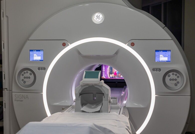
MRI is a slow imaging modality, where each series of images typically takes several minutes to acquire, a time during which the patient needs to lie still. The research group has two major research themes to deal with this issue. One is ultra-fast imaging such as NeuroMix, producing nine series with the most common MR contrasts in under 3 min.

The second theme is prospective (i.e. real-time) motion correction so that patients can move their heads freely during the MR examination with good diagnostic image quality. This is particularly important for MRI examinations of the pediatric brain, where our goal is to drastically reduce the use of general anesthesia, which is resource-intensive and not completely risk-free. It also leads to long waiting times for MRI and long MRI examinations.


KS foundation
KS Foundation


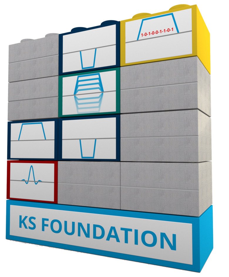
KS Foundation - Simplified pulse programming for faster development
 

To develop new advanced imaging techniques, and to aid collaboration between researchers, we have developed a simplified way to program MR systems from GE Healthcare, via an abstraction layer called KS Foundation. With this, pulse sequences can be written with significantly less code, and with a structure that enables more complex imaging techniques, such as NeuroMix. KS Foundation with associated pulse sequences is available to other MR researchers via ksfoundationepic.org, and is today used around the world.


Prospective (real-time) correction of head movements - Tracoline


Prospective (real-time) correction of head movements - Tracoline
Tracoline-system

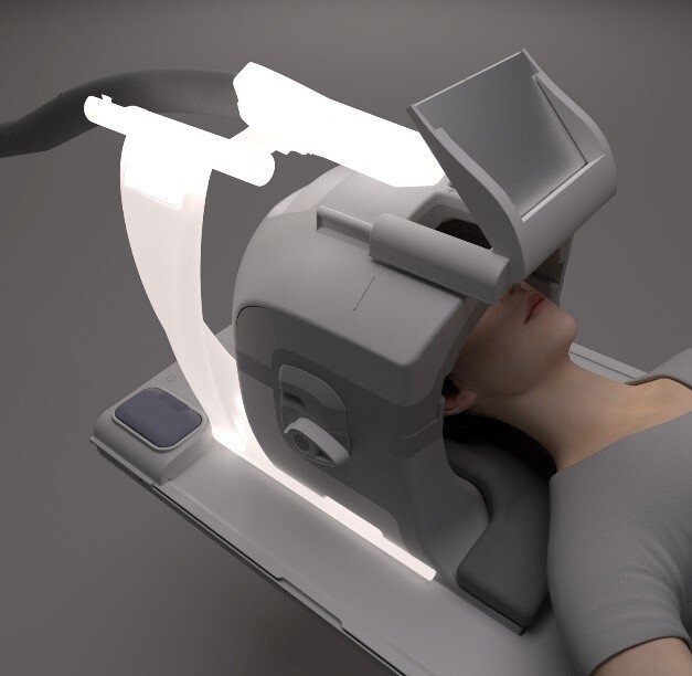
To make the MR image planes follow the patient's head a continuous measurement of the movement of the head is needed. For this purpose, we have purchased a fiber-optic infrared "point-cloud" camera (illuminated in the illustration) from a danish startup company. This device measures the position of the patient's head inside the MR scanner about 10 times/sec. That information is then communicated to the ongoing pulse sequence acquiring the current image (over several minutes).


scanner
A pediatric patient watches a film via mirror while the point-cloud camera, mounted near the head, records the patient's head movements.


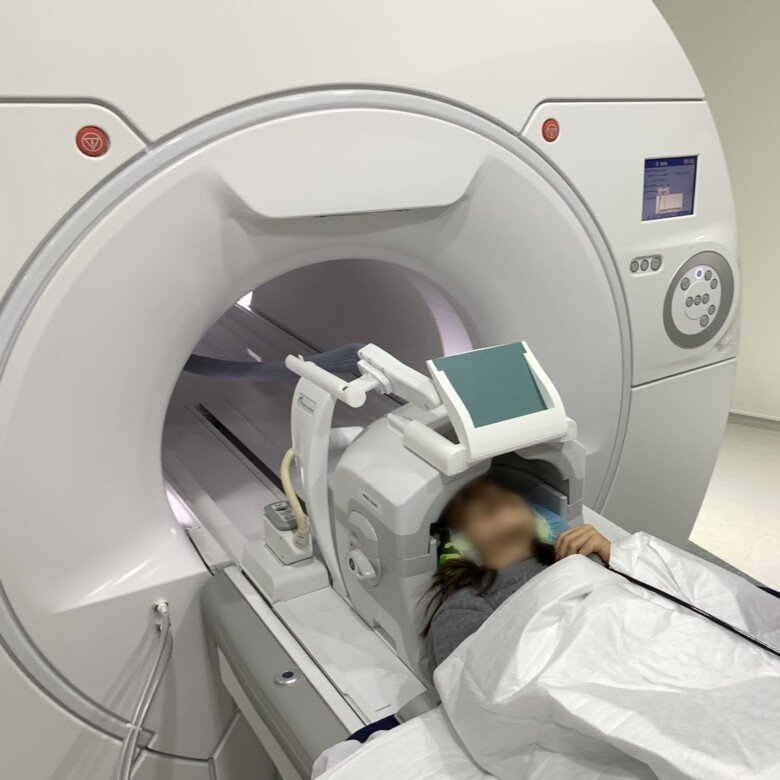
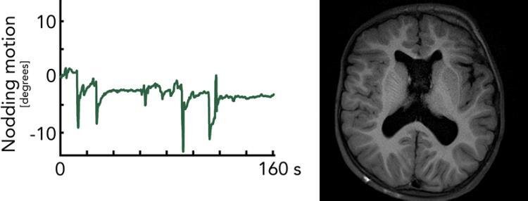
Motion monitoring of one of our pediatric patients being examined with our motion robust real-time corrected MR protocol based solely on our own pulse sequences. Above is a 3D rendering of the child's face as seen by the point cloud camera. Below is motion information for the entire MRI scan, which is continuously fed back to the imaging process to make the image planes follow the head movements


Prospective (real-time) correction of head movements - in-house developed device "WRAD"

We are also actively working on our own hardware-based sensor - referred to as a WRAD. This is an acronym for a Wireless Radiofrequency-triggered Acquisition Device. Put simply, it is a small wireless marker that synchronizes itself to the MRI scanner by detecting RF pulses. The device can then eavesdrop on the MR system’s gradient fields (typically very short navigators < 1 ms) and decode its location in the imaging coordinate frame to a high degree of accuracy. This allows us to perform low-latency (~3 ms) prospective motion correction with minimal modifications to the scanner setup. We believe the simplicity of this approach, with respect to workflow, is critical when performing prospective motion correction on real patients. With ongoing studies, including our sedation-free children’s brain protocol, we are constantly iterating the WRAD design with feedback from MR technologists and patients, but also from a generic need for better performance and stability. This project has a more “engineery” feel compared to some of our other goals and includes design, simulation, and 3D modeling with tangible-physical results.


nodding motion 2
The nodding (though-plane) part of the head motion was detected by WRAD during the T1FLAIR-PROPELLER acquisition of a 5Y pediatric patient. Despite repeated ~10 degrees of through-plane head motion, the image renders free of artifacts höger fri från bildstörningar.


Motion robust pulse sequences - Snapshot SWI-EPI


prospective motion correction

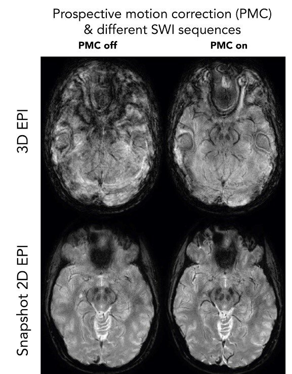
Despite real-time motion correction with Tracoline or WRAD, it is fundamentally difficult to obtain good image quality with long-TE 3D sequences such as SWI. Even with an ideal motion correction, the phase changes in the brain (which occur due to long echo times) result in image artifacts (top-right, 3D EPI). With our snapshot 2D SWI EPI, a complete image plane is obtained in 0.1 s under the same phase conditions in the brain. Several repetitions of the same image plane are averaged after motion correction to increase the SNR to the appropriate level.


Motion robust pulse sequences - Accelerated pseudo 3D PROPELLER

neuromix

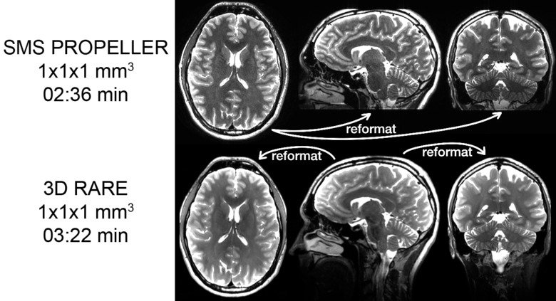
There is a clinical need for 3D RARE images with isotropic voxels that can be reformatted into several different planes, it increases the information for the radiologist and facilitates the review of the images. The sequences typically used to collect these 3D data sets require long echo trains, which makes them sensitive to movement and also leads to so-called "T2 blurring". In order to provide these reformatable volumes for patients who cannot lie still, we have developed a movement-robust alternative. A sequence that, through a combination of SMS acceleration, thin slices, and a PROPELLER acquisition, can obtain images with the same qualities as its 3D counterpart, but at the same time can withstand large head movements.


Ultrafast (and motion robust) MR - NeuroMix

neuromix

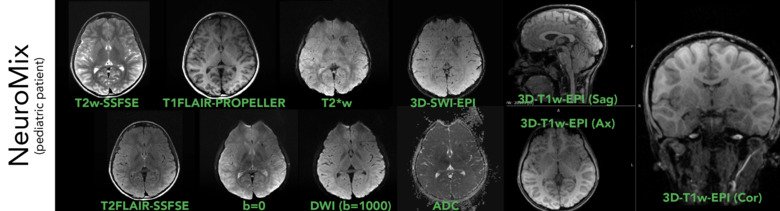
Here we show one slice per contrast from NeuroMix on a pediatric patient, as the first scan in our motion robust pediatric MR protocol. With a scan time for NeuroMix of ~2:40 min, where the 2D contrasts are motion robust and can be registered against each other in the reconstruction, these images usually turn out well even in the presence of larger head movements. And with future integration with WRAD & Tracoline, NeuroMix will be even more robust against head motion.


Publications

Selected publications


Niekerk A, Berglund J, Sprenger S, Norbeck O, Avventi E, Rydén H, Skare S. Control of a wireless sensor using the pulse sequence for prospective motion correction in brain-MRI. Magnetic resonance in Medicine 2022 Feb;87(2):1046-1061
View in Medline

View fulltext
10.1002/mrm.28994


Sprenger T, Kits A, Norbeck O, van Niekerk A, Berglund J, Rydén H, Avventi E, Skare S. NeuroMix - A single sequence brain exam. Magnetic resonance in Medicine 2022 May;87(5):2178-2193
10.1002/mrm.29120

View in Medline

View fulltext


Berglund J,  Sprenger T,  van Niekerk A,  Rydén H,  Avventi E,  Norbeck O,  Skare S. Motion-insensitive susceptibility weighted imaging. Magnetic resonance in medicine 2021 86;4 1970-1982

View in Medline

View fulltext

doi:10.1002/mrm.28850


Kits A,  De Luca F,  Kolloch J,  Muller S,  Mazya MV,  Skare S,  Delgado AF. One-Minute Multi-contrast Echo Planar Brain MRI in Ischemic Stroke: A Retrospective Observational Study of Diagnostic Performance. JOURNAL OF MAGNETIC RESONANCE IMAGING 2021 Oct;54(4):1088-1095 

View in Medline

View fulltext

doi:10.1002/jmri.27641


Ryden H,  Norbeck O,  Avventi E,  Skorpil M,  van Niekerk A,  Skare S,  Berglund J. Chemical shift encoding using asymmetric readout waveforms. MAGNETIC RESONANCE IN MEDICINE 2020 ; 1468-1480

View in Medline

View fulltext

doi:10.1002/mrm.28529


Berglund J,  Ryden H,  Avventi E,  Norbeck O,  Sprenger T,  Skare S. Fat/water separation in k-space with real-valued estimates and its combination with POCS. MAGNETIC RESONANCE IN MEDICINE 2020 83;2 653-661

View in Medline

View fulltext

doi:10.1002/mrm.27949


Norbeck O,  Sprenger T,  Avventi E,  Ryden H,  Kits A,  Berglund J,  Skare S. Optimizing 3D EPI for rapid T-1-weighted imaging. MAGNETIC RESONANCE IN MEDICINE 2020 84;3 1441-1455

View in Medline

View fulltext

doi:10.1002/mrm.28222


Avventi E,  Ryden H,  Norbeck O,  Berglund J,  Sprenger T,  Skare S. Projection-based 3D/2D registration for prospective motion correction. Magnetic resonance in medicine 2020 84;3 1534-1542

View in Medline

View fulltext

doi:10.1002/mrm.28225


Berglund J,  van Niekerk A,  Ryden H,  Sprenger T,  Avventi E,  Norbeck O,  Glimberg SL,  Olesen OV,  Skare S. Prospective motion correction for diffusion weighted EPI of the brain using an optical markerless tracker. MAGNETIC RESONANCE IN MEDICINE 2020 ; 1427-1440

View in Medline

View fulltext

doi:10.1002/mrm.28524


Rydén H,  Berglund J,  Norbeck O,  Avventi E,  Sprenger T,  van Niekerk A,  Skare S. RARE two-point Dixon with dual bandwidths. Magnetic resonance in medicine 2020 84;5 2456-2468

View in Medline

View fulltext

doi:10.1002/mrm.28293


Norbeck O,  van Niekerk A,  Avventi E,  Ryden H,  Berglund J,  Sprenger T,  Skare S. T-1-FLAIR imaging during continuous head motion: Combining PROPELLER with an intelligent marker. MAGNETIC RESONANCE IN MEDICINE 2020 ; 868-882

View in Medline

View fulltext

doi:10.1002/mrm.28477


Delgado AF,  Kits A,  Bystam J,  Kaijser M,  Skorpil M,  Sprenger T,  Skare S. Diagnostic performance of a new multicontrast one-minute full brain exam (EPIMix) in neuroradiology: A prospective study. Journal of magnetic resonance imaging : JMRI 2019 50;6 1824-1833

View in Medline

View fulltext

doi:10.1002/jmri.26742


Skare S,  Sprenger T,  Norbeck O,  Rydén H,  Blomberg L,  Avventi E,  Engström M. A 1-minute full brain MR exam using a multicontrast EPI sequence. Magnetic resonance in medicine 2018 79;6 3045-3054

View in Medline

View fulltext

doi:10.1002/mrm.26974


Norbeck O,  Avventi E,  Engström M,  Rydén H,  Skare S. Simultaneous multi-slice combined with PROPELLER. Magnetic resonance in medicine 2018 80;2 496-506

View in Medline

View fulltext

doi:10.1002/mrm.27041


Rydén H,  Berglund J,  Norbeck O,  Avventi E,  Skare S. T1 weighted fat/water separated PROPELLER acquired with dual bandwidths. Magnetic resonance in medicine 2018 80;6 2501-2513

View in Medline

View fulltext

doi:10.1002/mrm.27228


Engström M,  Mårtensson M,  Avventi E,  Norbeck O,  Skare S. Collapsed fat navigators for brain 3D rigid body motion. Magnetic resonance imaging 2015 33;8 984-91

View in Medline

View fulltext

doi:10.1016/j.mri.2015.06.014


Engström M,  Mårtensson M,  Avventi E,  Skare S. On the signal-to-noise ratio efficiency and slab-banding artifacts in three-dimensional multislab diffusion-weighted echo-planar imaging. Magnetic resonance in medicine 2015 73;2 718-25

View in Medline

View fulltext

doi:10.1002/mrm.25182


Skare S,  Hartwig A,  Mårtensson M,  Avventi E,  Engström M. Properties of a 2D fat navigator for prospective image domain correction of nodding motion in brain MRI. Magnetic resonance in medicine 2015 73;3 1110-9

View in Medline

View fulltext

doi:10.1002/mrm.25234


Engström M,  Skare S. Diffusion-weighted 3D multislab echo planar imaging for high signal-to-noise ratio efficiency and isotropic image resolution. Magnetic resonance in medicine 2013 70;6 1507-14

View in Medline

View fulltext

doi:10.1002/mrm.24594


Skare S,  Holdsworth SJ,  Lilja A,  Bammer R. Image domain propeller fast spin echo. Magnetic resonance imaging 2013 31;3 385-95

View in Medline

View fulltext

doi:10.1016/j.mri.2012.08.010


Engström M,  Bammer R,  Skare S. Diffusion weighted vertical gradient and spin echo. Magnetic resonance in medicine 2012 68;6 1755-63

View in Medline

View fulltext

doi:10.1002/mrm.24506


Members and contact

Group leader

All members of the group