Research - Jorge Ruas laboratory

Research in our laboratory is aimed at understanding the molecular mechanisms that mediate skeletal muscle adaptations to diverse challenges and their local and systemic consequences.

graphic illustration of ruas lab research focus

Sedentary lifestyles seem to come at a high cost to our health and have been linked to the incidence of several diseases including diabetes, obesity, cardiovascular disease, neurodegenerative diseases (such as Alzheimer’s and Parkinson’s), mood disorders (e.g. depression), and even cancer. Physical activity and skeletal muscle condition play a clear role in the prevention and treatment of these diseases. However, exercise programs are not always viable treatment options due to inherent disease characteristics such as muscle weakness, difficulty in movement, or, in particular, patient compliance.

By studying the mechanisms by which skeletal muscle adapts to different challenges and positively affects so many aspects of human health, we can learn valuable lessons that can be translated into future disease therapies.

Research Projects

The common goal of the projects being developed in our laboratory is to better understand the signal transduction and gene regulatory pathways that control skeletal muscle function in health and disease. With this knowledge, we aim at developing strategies that can be used as possible therapeutic avenues for the treatment of metabolic and degenerative diseases.

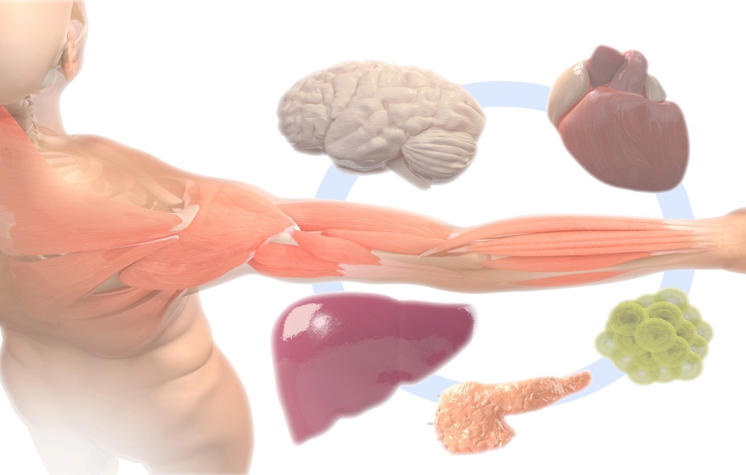
We are particularly interested in understanding how exercised or sedentary skeletal muscle can crosstalk with other organs, and how it can affect individual health and disease.

Mechanisms of regulation of skeletal muscle size and strength

Skeletal muscle is an extremely plastic tissue that can use energy to generate work, generate energy by breaking down proteins into amino acids, undergo atrophy, hypertrophy, and even change its metabolism when stimulated by distinct exercise programs (that is, endurance versus resistance training). By using a combination of genomics and proteomics approaches with different genetic models of skeletal muscle conditioning we hope to uncover new pathways important for the regulation of skeletal muscle mass and function.

Skeletal muscle metabolism and its crosstalk to the central nervous system

The beneficial effects of physical exercise on the prevention and treatment of neurodegenerative and mood diseases are well known. However, the mechanisms that mediate these effects are not completely understood. We have recently shown that skeletal muscle metabolism of the tryptophan metabolite kynurenine can protect form stress-induced depression by preventing the neuroinflammation associated with brain accumulation of this molecule. Our lab continues invested in trying to better understand how information is conveyed between peripheral tissues and to the brain.

A bioengineering approach to study inter-organ communication

Although it is clear that during skeletal muscle adaptation to diverse stimuli, including physical exercise, information is exchanged between this and other tissues in the body, the mechanisms that mediate this communication remain in great part elusive. Additionally, there are inherent difficulties in establishing causality in in vivo systems and the search for circulating secreted factors is hindered by the challenge of unbiased screening in the complex blood compartment. We are using mini-tissue microfluidics systems to model the information flow between skeletal muscle and other tissues. Tissue-specific functions are monitored by sensors including temperature probes and contractility measurements. The defined nature of these systems will facilitate the identification and manipulation of circulating factors affecting tissue function and metabolism.

graphic illustration of ruas lab research focus

Modulation of PGC-1alpha1 activity in skeletal muscle

Skeletal muscle adaptation to exercise training is mediated by the concerted actions of several transcriptional regulators, among which PGC-1alpha proteins play a central role. PGC-1alpha1 belongs to a family of coactivator proteins (together with PGC-1beta and PRC) and is highly induced in tissues with high energy demands by signals that increase energy output, such as cold and exercise. When activated, PGC-1alpha1 induces genes relevant to mitochondrial biogenesis, adaptive thermogenesis, lipid and glucose homeostasis, fiber-type switching, among other processes. For these reasons, deficiencies in PGC-1alpha1 activity have been suggested to be involved in pathogenic conditions such as obesity, diabetes, sarcopenia, and neurodegeneration. Conversely, it has been shown that overexpression of PGC-1alpha1 in murine skeletal muscle has several beneficial effects. We are developing strategies to increase PGC-1alpha1 levels in muscle, which small molecule technologies. The efficiency of these strategies is being tested in vitro and in vivo in the context of metabolic and muscular diseases.

PGC-1alpha splice isoform function in skeletal muscle

We have identified several new PGC-1alpha variants that are expressed at significant levels in skeletal muscle and several other tissues. Transcription of these isoforms is initiated at an alternative promoter of the PGC-1alpha gene, which seems to also induce alternative mRNA splicing. Among these, the PGC-1alpha4 variant is induced by resistance exercise training and specifically promotes skeletal muscle growth and strength. Importantly, transgenic animals with elevated PGC-1alpha4 levels in skeletal muscle show increased exercise performance, and resistance to atrophy and to cancer-induced cachexia. The functions of PGC-1alpha2 and 3 remain unknown. We are exploring the biological functions of these proteins using a combination of biochemical and genetic approaches.

Financial support

Current

Previous

JR
Content reviewer:
26-05-2023