Germ cell biology and developmental programming in epigenetic inheritance of diseases – Qiaolin Deng's research group

Reproduction is a fundamental process for passing traits from parents to offspring, traditionally believed to occur only through germline genetic modifications. However, evidence shows that early-life environmental exposures, influenced by parental health and developmental programming, can permanently impact offspring's health and disease risk. My lab studies how these interconnected processes, broadly termed as nature and nurture, shape the health trajectory of offspring.

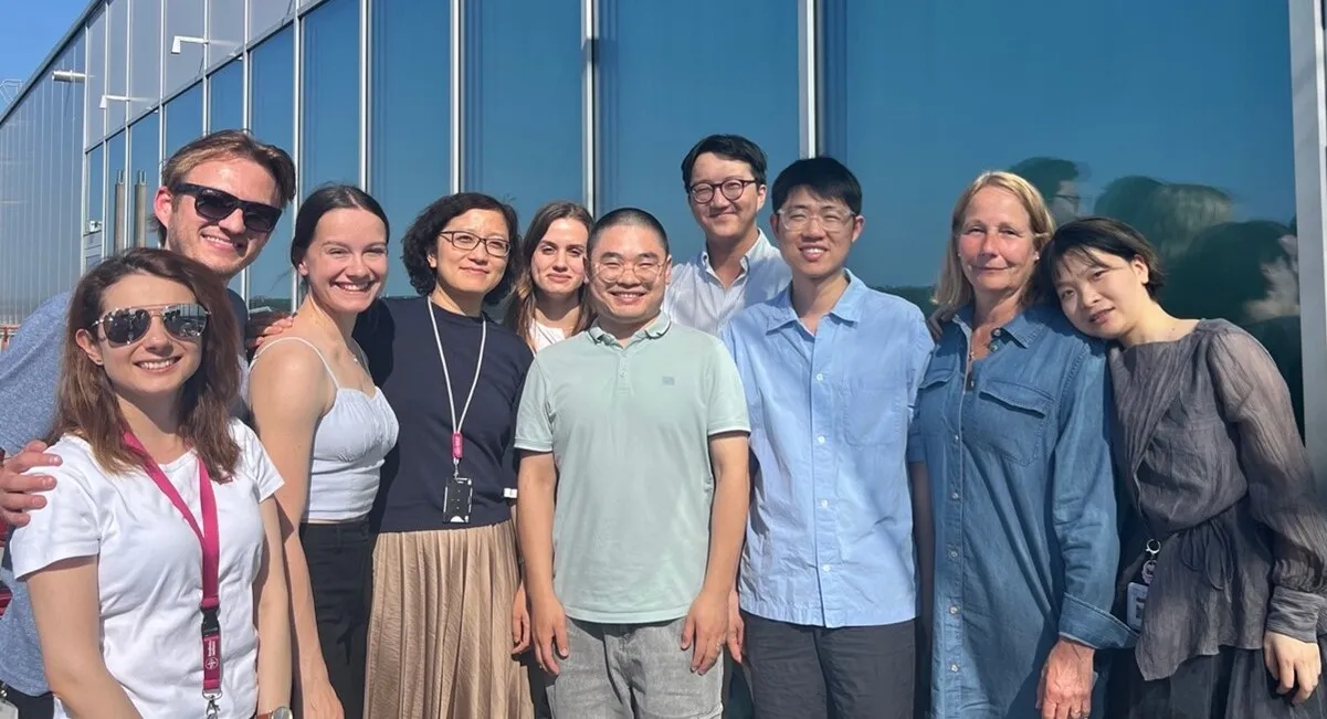
A group of people in front of a grey building
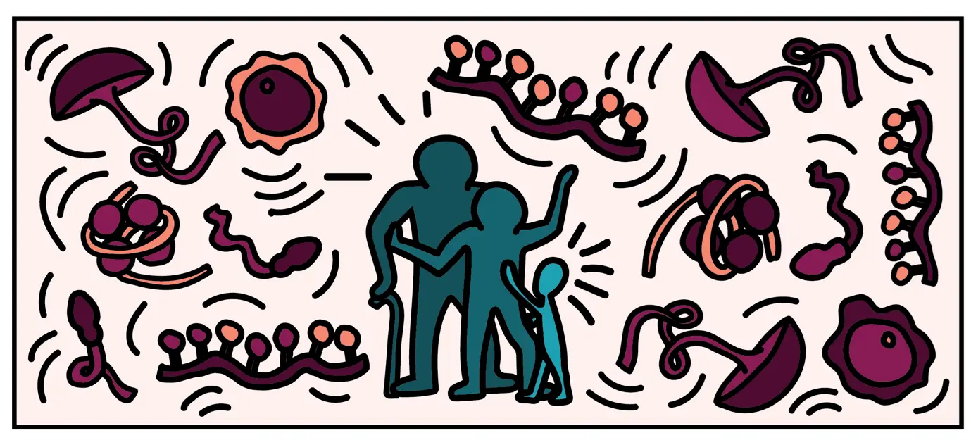
pink illustration with people and molecules
Illustration by Erin M. Slatery


Our research group is interested in the developmental principles of germline specification in health and disease using mouse models and human stem cell cultures coupled with state-of-the-art molecular and cellular tools. Moreover, we investigate how maternal diseases conditions impact the health outcomes of future offspring through the process called developmental programming by placentas and/or germline modulation, a process known as epigenetic inheritance of disease or developmental origins of health and disease (DOHaD). Our current research focuses on polycystic ovary syndrome (PCOS) and diabetes in women taking advantage of disease mouse models, human cohort samples, single-cell sequencing and disease-derived placental organoids in microfluidic co-culture system. 
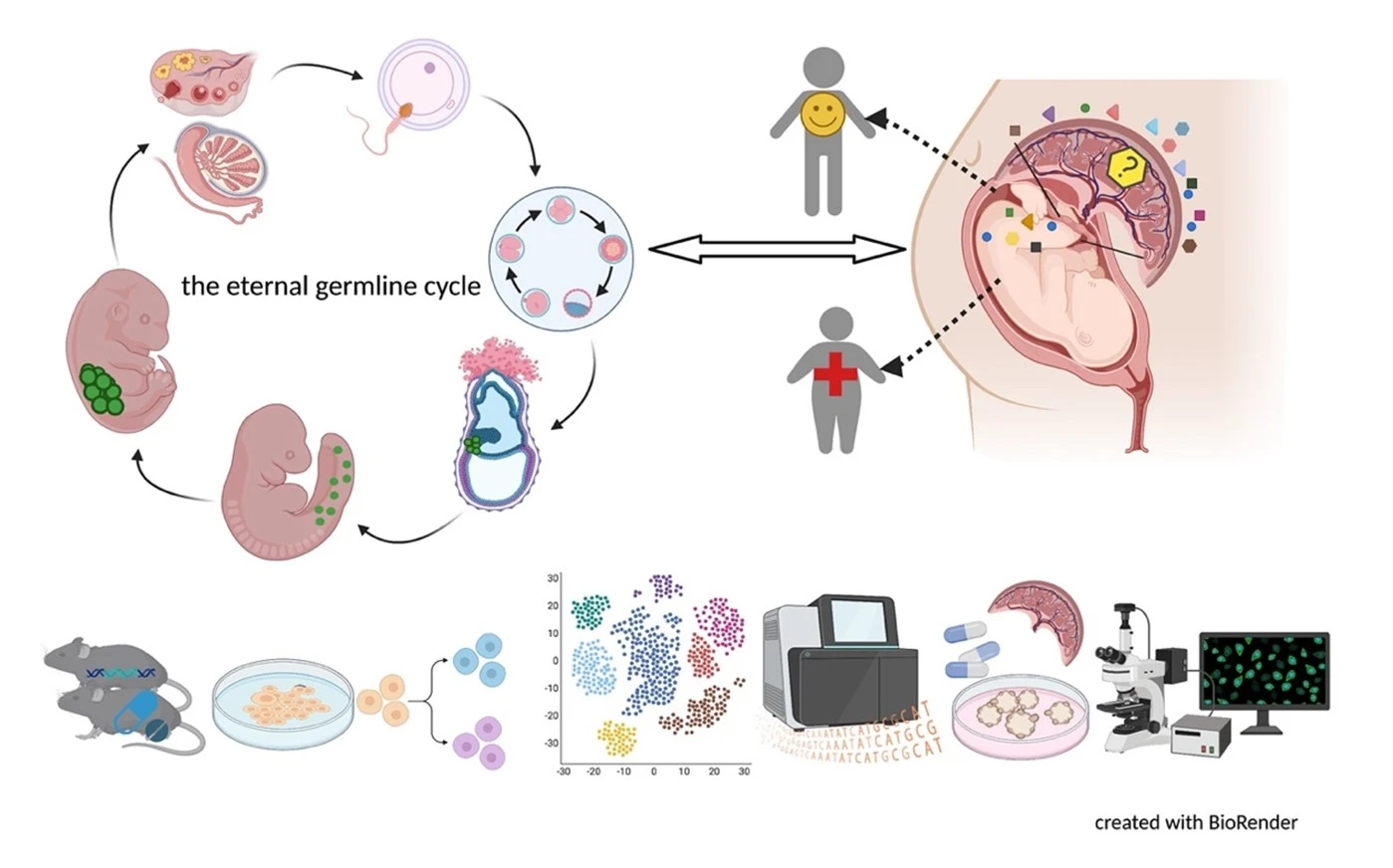
illustration of cells and fetus

Germ cells are often considered to be immortal, as they serve as the sole carriers of genetic and epigenetic information across generations, perpetuating life. The germline cycle is a lengthy process, beginning with early gastrulation and continuing through gametogenesis after birth. Any errors that occur during this process can have devastating and long-lasting effects. Recent advances in single-cell sequencing technology have greatly expanded our knowledge of germline development in mammals, but many questions remain unanswered. In this article, we aim to address some of these questions, such as (1) what regulates progenitor competence and how can we define cell quality for germline specification? (2) How is germline specification correlated with epigenetic remodeling including X-chromosome dosage effects? (3) How do certain genetic mutations affect gametogenesis?

Interestingly, there are two waves of epigenetic remodeling that occur after fertilization. These processes ensure the totipotency of the blastocyst for somatic lineage specification and establish a "clean slate" for germ cells, erasing any potentially harmful epigenetic modifications acquired from parents. These processes allow the organism to adapt to changing environmental conditions and minimize the risk of inheriting harmful traits. However, the completeness and faithfulness of these processes still need to be investigated. Increasing evidence shows that parental health conditions can predispose their offspring to develop diseases such as obesity, diabetes, cardiovascular disease, and behavioral disorders through developmental programming, a process called epigenetic inheritance of diseases. Mechanistic understanding of these processes is still sparse. We aim to answer questions such as (1) how do parental health conditions affect the germline, which further transmits phenotypic traits? (2) how does the placenta respond to adverse uterine environments, which can systematically modify the cellular and physiological functions of the developing fetues? (3) can we systematically model maternal disease signatures with offspring key organ signatures in humans using organoids and microfluidic coculture?

We are among those pioneers to apply and develop single-cell RNA sequencing (Smart-seq, Smart-seq2, LCM-seq etc). More tools to answer all these interesting questions are mouse disease models, human iPSC culture and differentiation, organoid culture, human sample cohorts and registry data together with other key cellular and molecular assays.

News

Publications

All publications from group members

Full list of publications

Full publication list @PubMed: https://bit.ly/4cq3qFO

Funding

Current and past funding

Karolinska Institutet  faculty-funded career position (total 4+2+5 years since 2015)

https://ki.se/en/about/faculty-funded-career-positions-and-consolidator-grant

Karolinska Instituet research grant

https://staff.ki.se/ki-research-grants-awards

Swedish Diabetes foundation project grant

https://www.diabetes.se/forskning/Diabetesfonden/

Swedish childhood diabetes foundation project grant

https://www.barndiabetesfonden.se/om-typ-1-diabetes/forskning

Swedish Research Council for Medical Research (VR) starting grant, project grant and consolidator grant

https://www.vr.se/

Wallenberg Academy Fellow

https://kaw.wallenberg.org/en/wallenberg-academy-fellows

Swedish Medicine Association starting grant

https://www.ssmf.se/

Åke Wibergs stiftelse

https://ake-wiberg.se/

Jeanssons stiftelser

https://jeanssonsstiftelser.se/

Staff and contact

Group leader

All members of the group

Visiting address

Karolinska Institutet, Physiology and Pharmacology, Biomedicum B5, Solnaväg 9, Stockholm, Stockholm, 17177, Sweden

Postal address

Karolinska Institutet, Physiology and Pharmacology, Biomedicum B5, Solnaväg 9, Stockholm, Stockholm, 17177, Sweden

Alumni

  • Han Hui-Pin (postdoc)
  • Ahmed Reda (postdoc)
  • Veeramohan Veerapandian (postdoc)
  • Menghan Wang (postdoc)
  • Shangli Cheng (postdoc)
  • Julio Agulia Benitez (postdoc)
  • Geng Chen (postdoc)
  • Yu Pei (PhD student)
  • Alice Larsson (master student)
  • Christina An Binh Nordentoft (master student)
  • Alisa Luo (biomedicine student)
  • Tom van Leeuwen (master student)
  • Hannah Schöler (master student)
  • Mathias Ahlqvist (master student)
  • Olli Ainasoja (master student)

Our collaborators

  • Elisabet Stener-Victorin, Dept. Physiology and Pharmacology, KI
  • Jan-Bernd Stukenborg, Dept. Women and Children’s health, KI
  • Martin Enge, Dept. Oncology and Pathology, KI
  • Kenny Rodriguez-Wallberg, Dept. Clinical Science, Intervention and Technology, KI
  • Sergiu-Bogdan Catrina, Dept. Molecular Medicine and Surgery, KI
  • Eva Hedlund, Dept. Biochemistry and Biophysics, Stockholm University
  • Xingqi Chen, Dept. Immunology, Genetics and Pathology, Uppsala University
  • Jun Wu, UT Southwestern Medical center, USA
  • Jiayu Cheng, School of Life Sciences, Tongji University, China
  • Weida Li, School of Life Sciences, Tongji University, China
  • Eszter Vanky, Institute for Clinical and Molecular Medicine, NTNU, Norway
  • Sandra Haider, Reproductive Biology Unit, Medical University of Vienna

Research projects

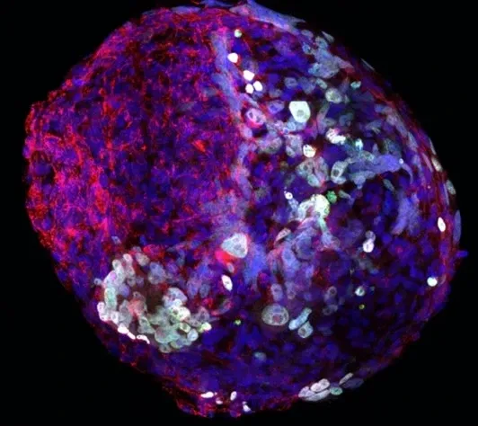
Microscope picture of a hPGCLC sphere culture
Sphere culture for hPGCLCs expressing TFAP2C (green), Mitotracher (red), DAPI (blue).

While specific projects may change over time, our dedication to the following research fields remains constant.

Research field I: Develop culture platform to differentiate germ cells towards maturation with epigenetic remodeling for the purpose of disease modeling and mechanistic understanding

The use of pluripotent stem cells for in vitro differentiation provides a most valuable approach to study human germ cell specification and to uncover genetic and epigenetic dysregulations associated with infertility. Our previous and recent work has demonstrated the establishment of formative pluripotency conditions for direct specification of primordial germ cell-like cells (PGCLCs) (Cheng et.al Cell Reports 2019, Luo et.al Cell Reports 2023). Building on this work, we plan to further develop in vitro differentiation platform using organoid co-culture to investigate the impact of genetic mutations on fertility.

Specifically, our research has shown focus on understanding the role of X-chromosome activity and mitochondrial dynamics in germ cell development.

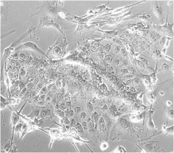
Microscope picture of a LT-hPGCLC coculture
Long-term (LT)-hPGCLC culture for 2 months with fetal gonadal somatic cells.

Research field II: Characterize the spatiotemporal regulation of germ cell migration and reveal the mechanisms of quality selection for gametogenesis

Germ cell migration is accompanied by extensive DNA demethylation and histone protein modification. We still know little about how these two sophisticated processes are coordinated. Furthermore, majority of migrating germ cells undergo apoptosis without further commitment to gametogenesis. We aim to use genetic mouse models, lineage tracing, single-cell sequencing among others to reveal gene function and mitochondrial dynamics during migration, epigenetic resetting, and meiosis entry. Especially, we are focusing on human genetic mutations implicated in infertility/subfertility.

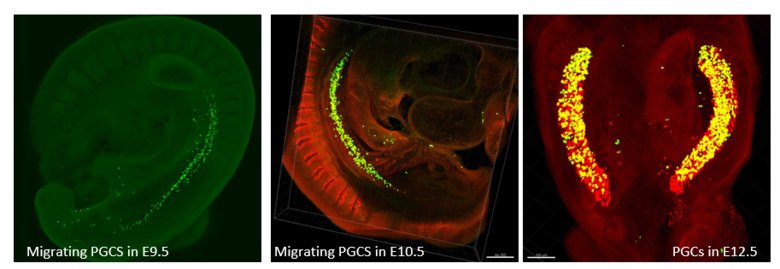
Three pictures in a row showing Migrating PGCS
Whole-mount immunostaining of PGCs. (A) Migrating PGCs in the E9.5 mouse embryo showing OCT4 staining. (B) Migrating PGCs in the E10.5 mouse embryo showing OCT4 and DPPA3 double positive staining. (C) PGCs in embryonic gonads in the E12.5 mouse embryo double positive for DPPA3 and XXX.
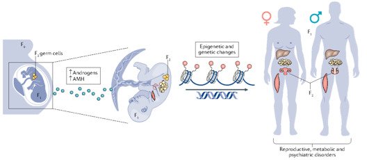
During pregnancy, environmental factors, such as exposure to maternal androgen or AMH excess adversely affect placental function and could result in epigenetic changes that affect the somatic and germ cells of the growing fetus. For example, histone modifications, DNA methylation and transcriptional regulation by non- coding RNAs might predispose both female and male offspring to develop reproductive and metabolic abnormalities as well as neuropsychiatric disorders. F0, the pregnant woman with PCOS; F1, her first-generation offspring; F2, her second-generation offspring. (Stener-Victorin E and Deng Q, Nat Rev Endocrinol 2021 Sep;17(9):521-533) Photo: n/a

Research field III: Investigate the role of germline transmission in epigenetic inheritance of diseases

Women are born with a pool of primary follicles, some of which are periodically matured for fertilization. Throughout a woman's reproductive life, the number and quality of the remaining follicles gradually decline, leading to a decrease in fertility and an increased risk of reproductive disorders. Besides genetics, environmental factors can affect the health and function of the oocytes through dynamic interaction with niche cells. We are particularly interested in how maternal endocrine diseases such as polycystic ovarian syndrome (PCOS) and type I diabetes impact transcriptome and metabolism of oocytes, which in turn affects their offspring health across generations independent of uterine environmental effects. We are using diseased mouse models, IVF and surrogacy, and single-cell sequencing together with phenotyping and molecular assays.  

Research field IV: Investigate the molecular basis of maternal-fetal communication in maternal endocrine diseases and their effects on offspring future health

Functional placentation and endometrial receptivity are essential to maintain a healthy pregnancy and influence fetal development. We have previously showed that reproductive and metabolic traits of maternal PCOS can be transmitted across generations (Risal, Pei et.al. Nature Medicine 2019). To further delineate the role of adverse uterine environment versus germline transmission, we are carrying on several projects to study the role of the placenta in developmental programming and offspring’s future health. As increasing evidence has shown that the placenta is not functioned as a passive barrier, instead actively responds and adapts to uterine environment to impact fetal development. It is important to elucidate molecular adaptation of placentas in maternal endocrine diseases in function to maternal clinical features including hormone profiles. We also aim to mediate the placental function and prevent the developmental programming. To address these questions, mouse disease models, single-cell sequencing of human placentas as well placental organoids based chemical screening are applied to systematically examine molecular and phenotypic traits of mother, placentas and offspring and build molecular link underlying the developmental programming effects.

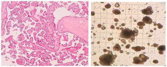
A: Human placenta section with H&E staining showing villous tree morphology. B: Human placental organoid culture
A: Human placenta section with H&E staining showing villous tree morphology. B: Human placental organoid culture.
Keywords:
Bioinformatics (Computational Biology) (applications, see 10610) Cell and Molecular Biology Developmental Biology Developmental Biology Diabetes Mellitus, Type 1 Endocrinology and Diabetes Epigenetic Memory Germ Cells Human Embryonic Stem Cells Hyperglycemia Induced Pluripotent Stem Cells Medical Biotechnology (focus on Cell Biology (incl. Stem Cell Biology), Molecular Biology, Microbiology, Biochemistry or Biopharmacy) Obstetrics, Gynaecology and Reproductive Medicine Organoids Physiology Placenta Polycystic Ovary Syndrome Single-Cell Analysis Show all
QD
Content reviewer:
30-10-2024