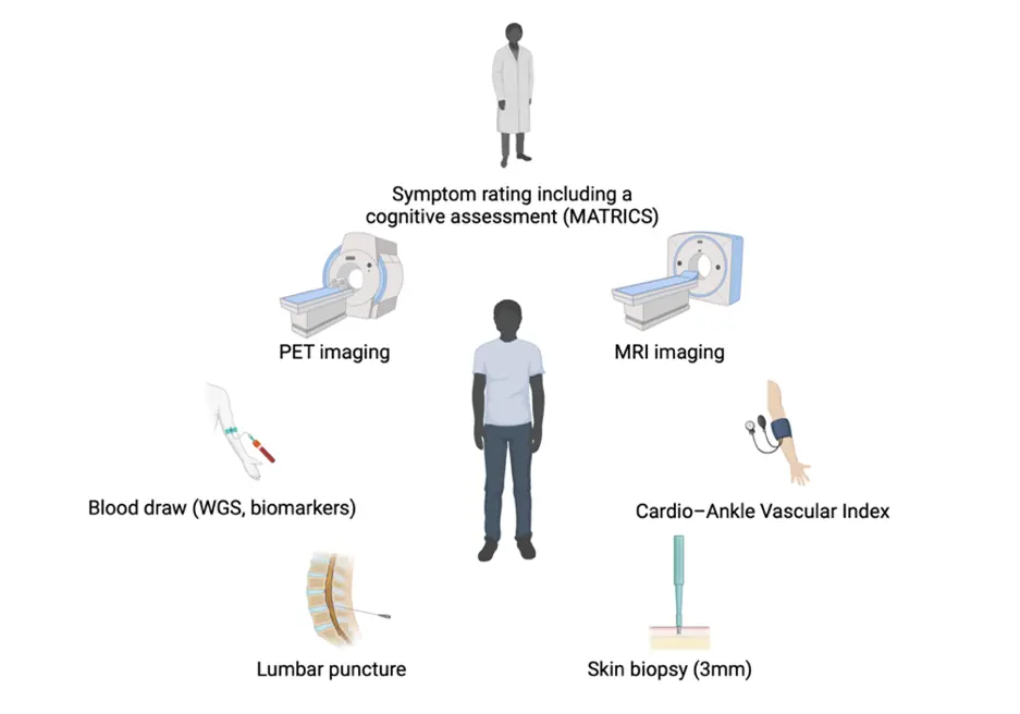
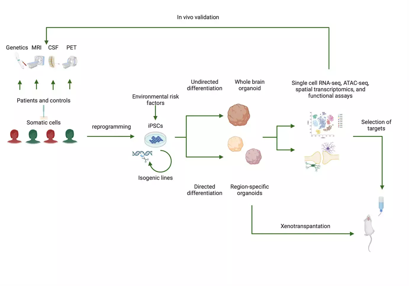
The laboratory is located in Biomedicum (Campus Solna) and uses models of the developing human brain (based on induced pluripotent stem cells) to study mechanisms that guide neurodevelopment and that are involved in different neurodevelopmental disorders (using genetic and environmental risk models). For this, we collect skin biopsies from patients at the psychiatric clinics in Stockholm and integrate experimental data with observational data obtained through genetic analyses, brain imaging, and analyses of cerebrospinal fluid.
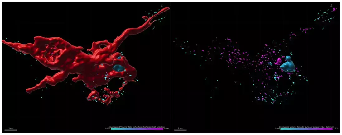
The clinical unit foremost works within the Karolinska Schizophrenia Project but also in collaboration with other cohort studies. Based on our earliest findings, we have a special interest in glial modulation of developing neuronal circuits and have developed brain organoid models that incorporates both microglia and oligodendrocyte lineage cells. Combined with different functional assays, single cell multiomics techniques are also commonly used in the laboratory to characterize brain organoids.
Projects
The current projects in our laboratory can be divided into four major categories:
- Assay development including cellular high-throughput approaches as well as more complete models such as organoids.
- Patient vs. control comparisons.
- Disease-orientated mechanistic studies using for example genetic engineering.
- High-throughput compound screening.
Group members
- Sanna Bruno, MD, PhD student
- Cristina Belotti, postdoc
- Asimenia Gkogka, PhD student
- Marja Koskuvi, postdoc
- Susmita Malwade, PhD student
- Lena Lundberg, Research nurse
- Martin Lundberg, MD, PhD student
- Ana Oliveira, postdoc
- Samudyata, PhD, senior lab manager
- Martin Schalling, Professor
- Ruth Schlissel, Research coordinator
- Alireza Salehi, PhD, affiliated to research
- Carl M. Sellgren, Associate Professor, Group leader
- Rose Temizer, PhD student
- Caroline Westerberg Hake, Research nurse
- JingJing Xu, PhD student
Research support
- Marianne och Marcus Wallenbergs Stiftelse
- Knut och Alice Wallenbergs Stiftelse
- Swedish Research Council
- StratNeuro
- Erling-Persson Foundation
- Hjärnfonden
- ALF
- SSMF
- One Mind Foundation
- Kaiser Permanente
- Karolinska Institutet
- Bjarne Ahlström’s Memorial Fund
Collaborations
Our research team works in close co-operation with several national and international groups in order to build a representative biobank of live cells from subjects with psychiatric and neurological disorders.
Our clinical projects are part of the Karolinska Schizophrenia Project (KaSP), a collaboration between the three principal investigators Carl Sellgren, Simon Cervenka and Sophie Erhardt. Our group is responsible for collecting somatic cells for reprogramming as well as to head the collection of clinical data together with the Cervenka Lab.
Selected publications
Cerebrospinal fluid concentration of complement component 4A is increased in first episode schizophrenia. Gracias J, Orhan F, Hörbeck E, Holmén-Larsson J, Khanlarkani N, Malwade S, Goparaju SK, Schwieler L, Demirel İŞ, Fu T, Fatourus-Bergman H, Pelanis A, Goold CP, Goulding A, Annerbrink K, Isgren A, Sparding T, Schalling M, Yañez VAC, Göpfert JC, Nilsson J, Brinkmalm A, Blennow K, Zetterberg H, Engberg G, Piehl F, Sheridan SD, Perlis RH, Cervenka S, Erhardt S, Landen M, Sellgren CM. Nat. Commun. 2022 Nov 3;13(1):6427.
SARS-CoV-2 promotes microglial synapse elimination in human brain organoids. Samudyata, Oliveira AO, Malwade S, Rufino de Sousa N, Goparaju SK, Gracias J, Orhan F, Steponaviciute L, Schalling M, Sheridan SD, Perlis RH, Rothfuchs AG, Sellgren CM. Mol Psychiatry. 2022 Oct;27(10):3939-3950.
Increased synapse elimination by microglia in schizophrenia patient-derived models of synaptic pruning. Sellgren CM, Gracias J, Watmuff B, Biag JD, Thanos JM, Whittredge PB, Fu T, Worringer K, Brown HE, Wang J, Kaykas A, Karmacharya R, Goold CP, Sheridan SD, Perlis RH. Nat Neurosci. 2019 Mar;22(3):374-385.
Neurodevelopmental disorders-high-resolution rethinking of disease modeling. Khodosevich K, Sellgren CM. Mol Psychiatry. 2023 Jan;28(1):34-43.
Contact
Carl Sellgren Majkowitz
Researcher;Assistant ProfessorRuth Schlissel
AssistantNews
 Photo: iStock
Funda Orhan wants to understand what causes schizophrenia
Funda Orhan tries to understand what causes schizophrenia. One of her driving forces it that her research may ultimately lead to helping people suffering from mental illness. Read the full article published on Hjärnfonden’s website on 29 March 2019 (in Swedish).
Links to news articles
Support our research
 Photo: Creative Commons CC0
Make a donation to our research
Your support means a lot to the success of research. This allows us to go further in our efforts to improve human health through research and education.