Introduction
Breast cancer is the most common cancer among women worldwide and remains a major cause of cancer-related mortality despite substantial advances in screening and treatment.
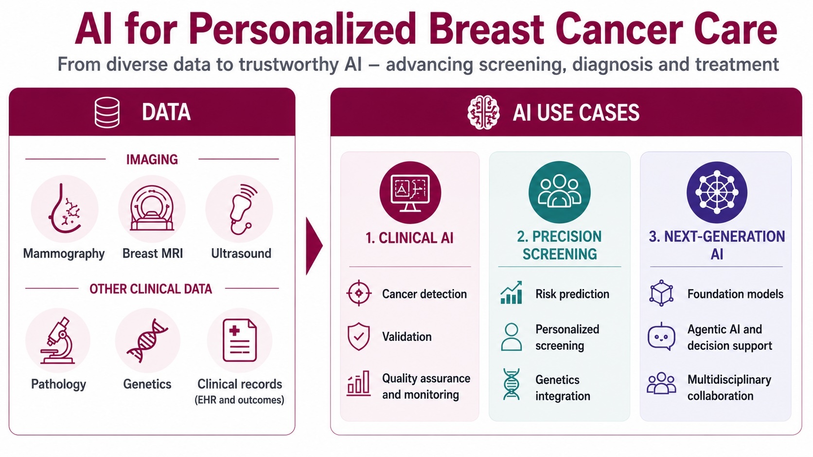
Our research explores how artificial intelligence can support more personalized, effective, and equitable breast cancer care. We develop and evaluate AI methods across the entire care pathway—from identifying women at increased risk of disease, to improving cancer detection in screening, supporting treatment planning, and enabling multidisciplinary decision-making.
A defining feature of our work is the integration of methodological development with prospective clinical evaluation. By combining expertise in radiology, computer science, epidemiology, oncology, and implementation science, we aim not only to develop new AI technologies, but also to understand how they can be safely, responsibly, and effectively implemented in clinical practice.
Research Focus
Clinical implementation of AI in breast screening
ScreenTrustCAD
To understand how artificial intelligence can be integrated into routine breast cancer screening, we lead the clinical study ScreenTrustCAD together with Capio S:t Göran Hospital. The study evaluates whether AI can safely replace one of the radiologists in a double-reading workflow and whether AI-supported screening can maintain cancer detection while reducing radiologist workload. In parallel, we collaborate with researchers in ethics and implementation science to better understand how radiologists and screening participants interact with AI systems in clinical practice.
Validation, fairness, and quality assurance of AI - VAI-B
Independent validation is essential for the safe implementation of AI in healthcare. Through the VAI-B project, we have established a collaboration between universities, healthcare regions, and patient organizations to evaluate AI algorithms in breast imaging. Current work focuses on algorithm performance across different patient populations, including disparities related to ethnicity and socioeconomic status, as well as methods for quality assurance and constancy testing of AI systems in clinical practice.
Precision screening and risk prediction
ScreenTrustMRI
Breast cancer screening should ideally be adapted to the individual woman. Together with collaborators at KTH, we have developed algorithms that estimate future breast cancer risk and the likelihood that an existing cancer is masked on mammography. These algorithms are being evaluated in the randomized clinical trial ScreenTrustMRI, which investigates whether risk-based MRI screening can reduce interval cancers and advanced cancers compared with standard mammography screening. The study also evaluates the health-economic implications of personalized screening strategies.
AI-based risk prediction and genetics - IMAxGENE
In the Swedish Cancer Society-funded IMAxGENE project, we combine mammography-based risk prediction with genetic risk information to improve individualized breast cancer risk assessment. The project aims to develop more accurate methods for identifying women at elevated risk and to explore how future screening and surveillance programmes can be tailored according to both imaging and genetic information.
Precision radiological diagnostics
Virtual Biopsy
We develop methods for prediction of tumour characteristics directly from imaging (“virtual biopsy”), and other quantitative MRI analyses. The purpose is not to completely replace real biopsies and histopathology, but rather to signal a potential need for renewed biopsy due to predicted tumor heterogeneity or temporal change in biological characteristics over time.
Cancer Segmentation
Our research increasingly focuses on multimodal breast radiology, including MRI and ultrasound. Therefore, we have had expert radiologists perform thousands of pixel-level annoations of examinations across all three modalities. We develop AI models for cancer and lymph node segmentation.
AI for predicting chemotherapy response in breast cancer – RadioVal
Within the Horizon Europe-funded project RadioVal, we participate in a multi-site European study developing and validating AI radiomics tools that predict preoperative therapy response. Our work spans the technical foundations and the clinical evaluation, where we coordinate a multidisciplinary reader study. By predicting treatment response earlier, the project aims to support more personalised therapy and reduce exposure to ineffective treatment.
Next-generation AI for breast cancer care
Generative AI for multidisciplinary cancer care – GenAI4Care
As part of the Horizon Europe project GenAI4Care, we collaborate with academic and clinical partners across Europe to develop and evaluate agentic AI systems that support multidisciplinary healthcare teams. The breast cancer use case focuses on helping clinicians synthesize information from imaging, pathology, clinical records, and treatment guidelines to support collaborative decision-making throughout the patient journey.
Evaluating foundation models for radiology – BenchmarkAI
Large vision-language models are rapidly transforming medical imaging. Through BenchmarkAI, a collaboration with the University of California, Berkeley, we are developing evaluation frameworks for foundation models in radiology. The project investigates how such models perform across a range of imaging tasks, including chest radiography, computed tomography, and magnetic resonance imaging, with the goal of establishing robust evaluation methods for future clinical applications.