Vascular morphogenesis and function in health and disease – Lars Jakobsson group

We are studying how blood and lymphatic vessels acquire and maintain their architecture and function in development and disease, aiming to improve therapies of vascular-connected pathologies. The research is directly related to vascular anomalies, cancer, heart disease, stroke, bleeding disorders, diabetic complications, eye disease, and oedema.

We are currently recruiting new team members! If you want to drive exciting projects as Postdoc, please contact: Lars Jakobsson via email: Lars.jakobsson@ki.se. Positions are fully funded.

Vascular morphogenesis and function in health and disease
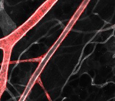
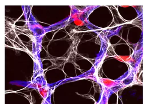
The vasculature of the skin. Mural cells (red) enclose ECs (white) of arteries and veins. Photo: Lars Jakobsson

Overview

Dysfunction and mispatterning of the blood and lymphatic vasculatures are central in the progression or development of several diseases such as cancer, lymphedema, diabetic retinopathy, trauma, stroke complications, hereditary haemorrhagic telangiectasia (HHT) and ischemic heart disease.

The formation and function of the vascular system of arteries, capillaries and veins rely on properties of the components of the vascular wall; the endothelial cells (ECs), the supportive mural cells (pericytes and smooth muscle cells), astrocytes (in the CNS) and their shared basement membrane (BM).

Aim

We study molecular and cellular processes whereby the blood and lymphatic vasculatures adapt their size, architecture, and function to optimally meet the changing demands from tissue. The aim is to utilize the acquired knowledge to develop means to prevent vascular malformation and malfunction in disease. The research field is directly relevant to a vast number of human pathologies, including vascular anomalies, heart disease, stroke, diabetes complications, cancer, lymphedema, and inflammatory diseases.

Technology and resources

  • Mice with inducible and cell specific genetic gain- and loss- of function in combination with a repertoire of conditional and cell specific reporters allowing for lineage tracing, clonal analysis and cell sorting.
  • Multi-photon intravital imaging
  • Confocal live cell imaging
  • Fluidics and in vitro experimentation
  • Tissue-clearing techniques
  • Light-sheet microscopy
  • scRNA-sequencing
  • Large scale proteomics
  • Mouse embryonic stem cell models
  • AAV-mediated gene modulation

Articles about the group

Research projects

The vasculature of the developing retina. Endothelial cells (blue), pericytes (red) and astrocytes (white) interact to form the functional vasculature.
The vasculature of the developing retina. Endothelial cells (blue), pericytes (red) and astrocytes (white) interact to form the functional vasculature. Photo: Lars Jakobsson

Introduction


Our work has exposed fundamental principles of how endothelial and mural cells organise into functional arteries, capillaries and veins through sprouting angiogenesis. We now know that this involves extensive cellular dynamics instructed by signalling via serum- and tissue- derived cytokines, inter-endothelial communication as well as flow-mediated shear forces. We deepen the knowledge on the interplay between, and impact of, several signalling cascades that are central for creation and maintenance of vessel architecture and function. By applying state-of-the-art basic science to resolve clinical problems, we aim to advance treatment of several vascular-related diseases. Please see our publications for details on previous contributions.

Mosaic gene
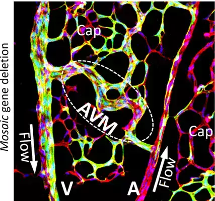
Picture legend: The retinal vasculature of a genetically modified mouse allowing for inducible mosaic endothelial cell-specific deletion of endoglin, thereby modelling human vascular disease. An abnormal shunt (arteriovenous malformation) directly connects the artery (right) and vein (left) thereby bypassing the normal capillary bed. The tissue is immunolabelled to identify endothelial cells (red/yellow) and their nuclei (blue). Photo: Yi Jin/Jakobsson

Mechanisms of vascular malformation

Endoglin is an EC expressed coreceptor for bone morphogenetic protein-9/10, a ligand found in serum. Loss-of-function mutations in the endoglin gene cause the human disease HHT1, also known as the Osler Weber Rendu syndrome, characterised by vascular abnormalities such as arterio-venous malformations that lead to bleeding, anemia and high output heart failure. Recent advances have provided promising treatments but with relapse and side effects. We have shown that ECs require endoglin in order to respond correctly to blood flow. Without endoglin – such as in the disease HHT1 – ECs cannot control their size, migratory behaviour or proliferation in accordance with flow. We continue to dissect mechanisms of this phenomena utilizing unique genetic disease models applying tools mentioned above. We are now investigating new targets to treat HHT1.

Visualizing the 3D architecture of the brain vasculature. Drug-induced endothelial specific endogenous fluorescence allows for 3D rendering of the brain vasculature. The tissue has been optically cleared and imaged using our multiphoton confocal system.
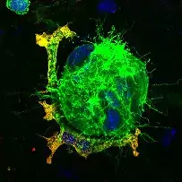
Modelling retinopathy of prematurity. Pathological angiogenesis of the retina occurs as a consequence of hypoxia (low oxygen). Dysfunctional microvascular tufts (spheres of dividing endothelial cells, green) form and are tightly associated with macrophages (red/yellow). Cell nuclei are in blue. Photo: Lars Jakobsson

The Basement membrane – facilitating structured signalling

ECs and pericytes rest on a matrix of several proteins that interconnect to form the basement membrane (BM). Laminins are key components of the vascular BM that transmits information to ECs mainly via the integrins. Alterations in the BM composition and organisation is commonly seen in tumours and vascular eye disease. Using conditional alleles, we study the role of laminins in establishment and function of the vasculature in physiology and pathology, such as during tumour growth and inflammation.

The vasculature in eye disease – a complete picture

Each compartment of the eye is supported by its individual specialised blood vasculature. These individual vascular beds are in turn involved in specific forms of eye disease. To extract deep information on structural properties of these vasculatures and their interplay in development, health and disease we refine and apply high resolution microscopy, including light-sheet fluorescence microscopy on intact murine eyes. These technologies allow for reconstruction of the whole eye in three dimensions, see 3D rendering below.

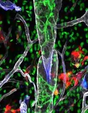
Lymphatic collecting vessels of the ear. The central part of the left image visualises a collecting valve. The colours indicate certain cellular subtypes or matrix proteins. Photo: Lars Jakobsson

Patterning and transformation of the lymphatic vasculature

The lymphatic vasculature drains fluid from tissue, via its blind-ended capillaries through collecting vessels to blood circulation. We have shown that recruitment of smooth muscle cells to collecting vessels relies on the secretion of PDGFB from lymphatic endothelial cells, in turn required for their contractile function. The importance of lymphatic vascular functionality is highlighted by extensive oedema in specific genetic diseases affecting lymph valves, as well as following lymph node removal in breast cancer.

Here we study specific genes and signalling cascades that have been associated with rare transitions of the lymphatic endothelium into cancers. Through careful analysis of the progression in experimental models we hope to find key features that can be modulated as part of future therapy.

Publications

Selected publications