Developmental biology and regenerative medicine – Igor Adameyko's research group

We look at a range of live systems from a mechanistic point of view, trying to reverse engineer different aspects of multicellular life. Tracing the incremental advancements in development of multicellular organisms from a single cell perspective allows better understanding of the complexity of the entire organism or organ system in a final phase.

An illustration from Igor Adameyko lab
Copyright: Igor Adameyko lab

Our Research

The knowledge gained from developmental biology research is widely applied in regenerative medicine. Thus, we hope to improve human health via discovering new fundamental ideas about how development, stem cells, and regeneration work.

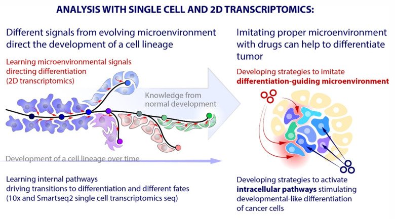
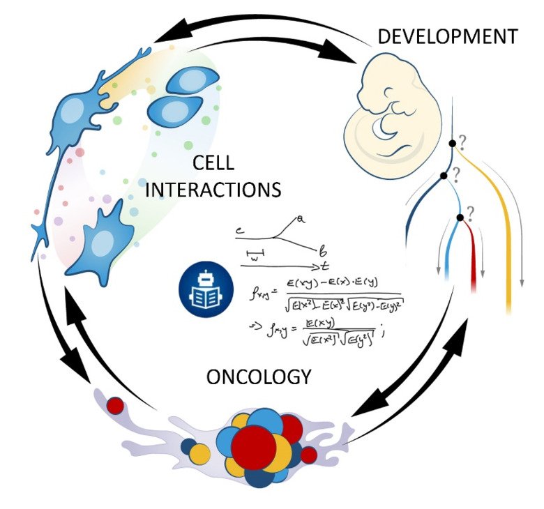
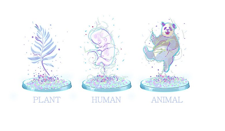
Our laboratory advances a broad spectrum of projects related to developmental biology, stem cells, EvoDevo and regenerative medicine. The methodology includes classical developmental biology approaches blended with single cell transcriptomics, 2D sequencing and 3D-reconstructions of tissues and organs based on optical or X-ray methods (micro-CT, synchrotron).

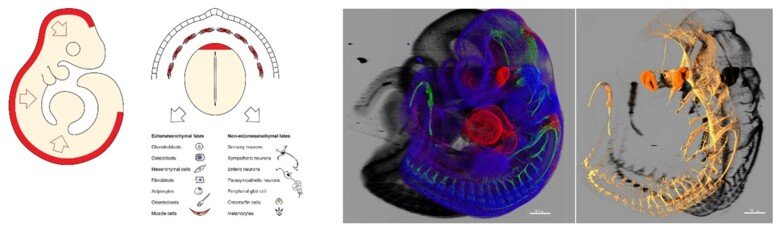
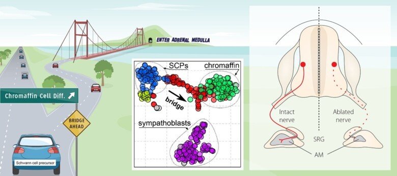
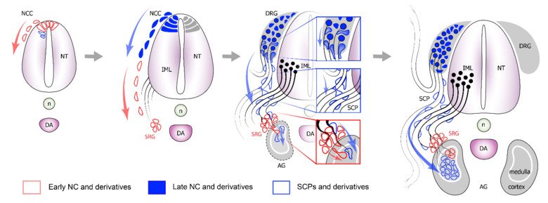
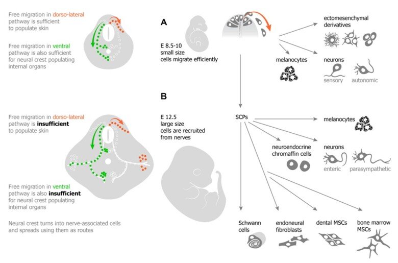
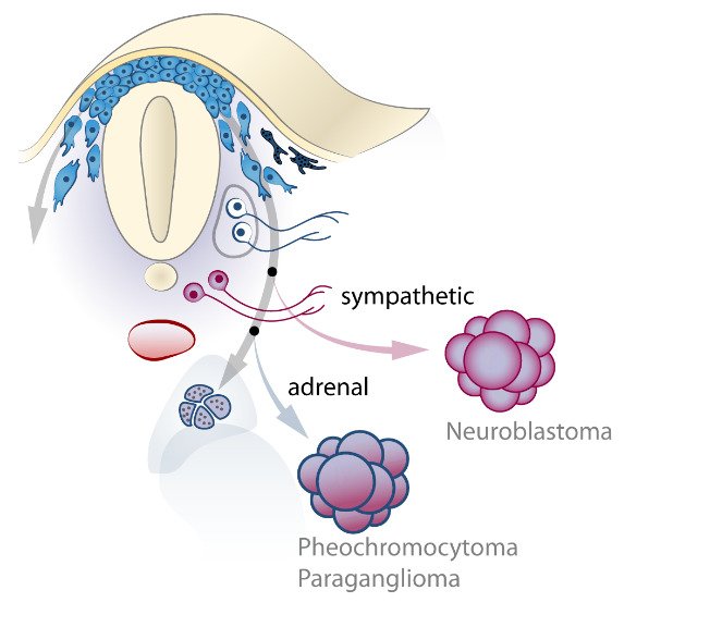
The neural crest stem cells is our primary model system, where we address general principles of cell fate choice, transcriptional and epigenetic control of a lineage progression, morphogenesis, and tissue shaping.

News from the group

Research projects

The conundrum of the neural crest multipotency and the mechanisms of a fate choice

Lately, the neural crest cells attracted a lot of attention, as these cells are multipotent and contribute to numerous cell and tissue types in the vertebrate embryo.

Neural crest cells generate skeletogenic tissues in our faces, the outflow tract of the heart, pigmentation, the autonomic and enteric nervous system, dental pulp and hard matrix of our teeth. These cells generate neurons that sense and convey touch, pain, heat and information about the position of the body parts in space, and many more. Because of their major role, neural crest cells are considered to be the 4th germ layer.

Although the multipotency of neural crest cells has been well known for decades, there is still an unsolved conundrum related to how cells make decisions and choose fate during embryonic development. For instance, currently, there is no genealogical model that would describe the multipotency type (individual or collective) in the neural crest lineage.

We are attempting to create such a model, to discover how neural crest cells choose fates during developmental progression towards multiple derivatives, and what defines the differences in such choices in the cranial and trunk regions.

Recently, we suggested a completely novel generalizable model of cell fate choice that we published in Science in 2019 (Soldatov et al., Science 2019). This model is based on competition between opposite genetic programs activated by the external environment in the same cells, followed by the eventual resolution of such conflicts with developmental timing. Indeed, cells always make such decisions during embryonic development, cancer progression, and in situations where they are responding to some form of stimulation.

The figure shows how migratory neural crest cells populate the E9.5 mouse embryo. Copyrights: Adameyko Lab.

Although we have generated a number of molecular insights into related mechanisms, we still lack mechanistic knowledge about how cells process information. This is our biggest question, and we are trying to understand what is it, that makes a decision right or wrong?

We have developed peculiar ideas about faulty plasticity, when cells cannot decide their own fate, which pre-conditions them for tumorigenesis. This is in many ways linked to the question of where cancer originates from.

Today, we are aware of many mutations and landscapes that promote cancer or help cancer cells to go beyond all the checkpoints. At the same time, we do not understand what is the fundamental driver of tumor cell identity during cancer initiation. In our lab, we work on these questions.

Key publication

Spatiotemporal structure of cell fate decisions in murine neural crest.
Soldatov R, Kaucka M, Kastriti ME, Petersen J, Chontorotzea T, Englmaier L, et al
Science 2019 Jun;364(6444):

Multipotency of SCPs - Role of SCPs in development of sympathoadrenal system

Recently, we have discovered an entirely new phenomenon in developmental biology – targeted recruitment and subsequent differentiation of multipotent cells from the pervasive peripheral nerves.

In brief, during recent years, one of the aims of our research was to define the source of the late embryonic and adult neural crest to harness the regenerative power of this cellular population.

With the help of various methods, we revealed a new principle allowing embryos to position cells during histogenesis: nerve-associated glia and neural crest-like progenitor pools undergo nerve-regulated migration and differentiation into peripheral autonomic neurons (Dyachuk et al., Science 2014), neuroendocrine chromaffin cells (Furlan et al., Science 2017), melanocytes (Adameyko et al., Cell 2019) and different types of mesenchymal cells (Kaukua and Khatibi et al., Nature 2014).

Our first breakthrough came in 2009 (Cell) and followed in 2012 (Development) demonstrating that Schwann cell precursors residing along growing peripheral nerves give rise to melanocytes (pigment cells in skin).

Today, many biological questions in the lab are focused around the idea that peripheral nerves are essential for the development of organs; they contribute multipotent precursor cells to local tissues and serve as delivery routes for universal progenitors giving rise to numerous cell types.

We apply single cell transcriptomics, color multiplexing in genetic tracing as well as functional studies in mouse, chick and zebrafish to address the relationships and molecular similarities between peripheral glial cells and migratory neural crest populations.

Our single cell approach coupled to powerful validation systems allows investigating gene regulatory networks that drive differentiation of the neural crest and nerve-associate multipotent Schwann cell precursors.

Figures illustrating the multipotency of SCPs. Role of SCPs in development of sympathoadrenal system.
Copyrights: Adameyko Lab.
Figures narrating a concept of how Schwann cell precursors (SCPs) give rise to chromaffin neuroendocrine cells of adrenal medulla.
The figure narrates a concept of how Schwann cell precursors (SCPs) give rise to chromaffin neuroendocrine cells of adrenal medulla. Copyrights: Adameyko Lab.

We recently explored sympathoadrenal development and discovered new Schwann cell precursor-dependent cellular origin of chromaffin cells using single cell transcriptomics (Furlan et al. Science 2017). Next, we started to investigate the malignancies arising from the sympathoadrenal origin such as neuroblastoma, pheochromocytoma and paraganglioma to relate intra-tumor heterogeneity to the potential cell of origin.

In our preliminary data, we observed that these tumors are highly heterogeneous and resemble stages of the neural crest differentiation into chromaffin, glomus, or sympathetic cells. Clarifying the role of the nerve-associated Schwann cell precursors in the development of sympathoadrenal organs and corresponding malignancies might help to reveal the causes of disease heterogeneity.

Creating “comparative identity maps” using single cell analyses of tumors and healthy progenitor populations will allow us to identify the precise developmental and adult origin cell types and corresponding differentiation statuses for different subtypes of neural crest tumors.

Figures summarizing the role of Schwann cell precursors in vertebrate embryonic development.
The figure summarizes the role of Schwann cell precursors in vertebrate embryonic development. Copyrights: Adameyko Lab.

Key publications

Multipotent peripheral glial cells generate neuroendocrine cells of the adrenal medulla.
Furlan A, Dyachuk V, Kastriti ME, Calvo-Enrique L, Abdo H, Hadjab S, et al
Science 2017 07;357(6346):

Neurodevelopment. Parasympathetic neurons originate from nerve-associated peripheral glial progenitors.
Dyachuk V, Furlan A, Shahidi MK, Giovenco M, Kaukua N, Konstantinidou C, et al
Science 2014 Jul;345(6192):82-7

Nerve-associated neural crest: peripheral glial cells generate multiple fates in the body.
Petersen J, Adameyko I
Curr Opin Genet Dev 2017 Aug;45():10-14

Returning neural crest-derived tumors back onto developmental path

Some malignant tumors in the human body originate from cells of the neural crest lineage. Those include melanoma, neuroblastoma, pheochromocytoma, schwannoma and many others.

Figures illustrating the knowledge flow between developmental biology approach and cancer heterogeneity-based extermination of tumors.
The figure suggests the knowledge flow between developmental biology approach and cancer heterogeneity-based extermination of tumors. Copyrights: Adameyko Lab.
Figures illustrating Returning neural crest-derived tumors back onto developmental path.
Copyrights: Adameyko Lab.

At the moment, we are aware of few productive attempts to differentiate tumors instead of killing them and thus subjecting cancer cells to further evolution. At the same time, a systematic approach to devise such strategies is currently missing. For many tumors we don’t know how to identify inter-cellular signals that could be either disrupted or induced (directly or by mimicking their downstream effects) that would push tumor cells towards differentiation.

Figures illustrating Returning neural crest-derived tumors back onto developmental path. Development, cell interactions, oncology.
Copyrights: Adameyko Lab.

Similarly, signaling interactions between different components of the tumor microenvironment and their interactions with the tumor cells themselves remain poorly characterized, despite numerous focused efforts. Thus, as a constellation of four different laboratories supported by the ERC Synergy project, we aim to develop and test a novel systematic approach for identifying recurrent cell-cell interactions, and apply it to find new ways to differentiate highly malignant tumors, preventing their metastasis.

The neural crest cancers offer a compelling test case as they often represent examples of incomplete differentiation. This approach can be, of course, combined with chemotherapy and surgery-based tumor-eliminating strategies.

The enigma of human facial diversity 

Another large-scale project we are working on is focused on understanding the molecular mechanisms behind human facial diversity. We humans are the only animals that communicate identity via individual-specific variation of facial shape.

In this respect, humans are unique. Other animals recognize each other rather based on the general appearance, smell, posture, behavior, coloration of fur and feathers, etc. Here we make an attempt to discover the genetic and molecular basis of such healthy facial shape variation.

Overall, we are trying to understand what makes us different, unique, and beautiful. Because our faces develop in utero from neural crest stem cells, these multipotent progenitors become the center of our study. We investigate a variety of the developmental aspects involved in facial shaping by cranial neural crest together with comparative genomics and transcriptomics in humans and animal models.

Figures illustrating craniofacial skeletal arrangements in various mutated mouse embryos.
The figure shows craniofacial skeletal arrangements in various mutated mouse Copyrights: Adameyko Lab.

Publications

Signals from the brain and olfactory epithelium control shaping of the mammalian nasal capsule cartilage.
Kaucka M, Petersen J, Tesarova M, Szarowska B, Kastriti ME, Xie M, et al
Elife 2018 06;7():

Oriented clonal cell dynamics enables accurate growth and shaping of vertebrate cartilage.
Kaucka M, Zikmund T, Tesarova M, Gyllborg D, Hellander A, Jaros J, et al
Elife 2017 04;6():

The Nervous System Orchestrates and Integrates Craniofacial Development: A Review.
Adameyko I, Fried K
Front Physiol 2016 ;7():49

Evolution of cell types and mechanisms controlling cell identity

The group also works on a problem of evolution of cell types and animal designs that result from cell type evolution. As cells became more and more diverse in organisms over geological time, they built bodies that are more sophisticated, by assigning new functions and properties to biological tissues. All great evolutionary leaps and transitions in animal body plans presumably involved elaboration of novel embryonic cell types – for instance, specific classes of embryonic progenitors.

Figures illustrating evolution of cell types and mechanisms controlling cell identity.
Copyrights: Adameyko Lab.

To be more detailed, we are interested in the neural crest evolution and the logic of expansion of the plethora of neural crest-derived fates in different chordate animals. Based on our neural crest-related research, we aim to generalize the molecular principles of cell type diversification and division of labor. Today, we apply comparative single cell transcriptomics to compare cell phenotypes and identities from different animals and to track how the cells change over geological time.

Publication

Prototypical pacemaker neurons interact with the resident microbiota.
Klimovich A, Giacomello S, Björklund Å, Faure L, Kaucka M, Giez C, et al
Proc Natl Acad Sci U S A 2020 07;117(30):17854-17863

The progeny of the neural crest builds our teeth

Mammalian teeth are built of many cell types including those producing hard matrix – odontoblasts making dentin, ameloblasts producing enamel and cementoblasts generating tooth cement.

Numerous cell subtypes reside inside of the soft dental pulp and in the surrounding tooth follicle. Vessels and sensory nerves complete the structure of a tooth. Many of these dental cells are neural crest-derived and commit to dental-specific fates because of the crosstalk between the oral epithelium and the neural crest-derived mesenchyme, resulting in a forming tooth germ.  

For instance, during this process, the exact specification and differentiation of the odontoblast lineage has not been fully clarified, and the eventual fating of mesenchymal neural crest progenitors was not fully addressed.

As a part of collaboration with laboratories of Kaj Fried and Jan Krivanek, we currently work on the heterogeneity of cell populations building our teeth, aiming to identify region- and tooth type-specific stem cells and define novel regenerative strategies. Recently, we utilized single cell transcriptomics to address the cell type heterogeneity in human and mouse teeth (please see Krivanek et al., Nature Communications 2020).

In that study, we reported an unappreciated cellular complexity of the continuously-growing mouse incisor, which suggests a coherent model of cell dynamics enabling un-arrested growth. This model relies on spatially-restricted stem, progenitor and differentiated populations in the epithelial and mesenchymal compartments underlying the coordinated expansion of two major branches of pulpal cells and diverse epithelial subtypes.

Further comparisons of human and mouse teeth yield both parallels and differences in tissue heterogeneity and highlight the specifics behind growing and non-growing modes. Despite being similar at a coarse level, mouse and human teeth reveal molecular differences and species-specific cell subtypes suggesting possible evolutionary divergence.

Today, we continue this work and attempt to investigate phenotypic plasticity of cell types within teeth resulting from regenerative or infection-related stimuli.

Figure revealing the diversity of dental cell types in a continuously growing mouse incisor.
The figure reveals the diversity of dental cell types in a continuously growing mouse incisor. Copyrights: Adameyko Lab.

Publications

Glial origin of mesenchymal stem cells in a tooth model system.
Kaukua N, Shahidi MK, Konstantinidou C, Dyachuk V, Kaucka M, Furlan A, et al
Nature 2014 Sep;513(7519):551-4

Heterogeneity and Developmental Connections between Cell Types Inhabiting Teeth.
Krivanek J, Adameyko I, Fried K
Front Physiol 2017 ;8():376

Dental cell type atlas reveals stem and differentiated cell types in mouse and human teeth.
Krivanek J, Soldatov RA, Kastriti ME, Chontorotzea T, Herdina AN, Petersen J, et al
Nat Commun 2020 09;11(1):4816

Other projects

We, as a team, support the diversity in thinking and favor natural curiosity of our members. This is why we always run some small hobby projects on a side in any area of modern biology and biomedicine – spanning from the mechanisms of integration operating within coral colonies to the studies of the composition of early multicellular life forms.

We believe that the lab environment must be invigorating, and the broad scope together with great opportunities to tackle natural puzzles serve this function pretty well.

Figures illustrating hobby projects at the Adameyko lab spanning from the mechanisms of integration operating within coral colonies to the studies of the composition of early multicellular life forms.
Copyrights: Adameyko Lab.

Publications

Selected publications

Members and contact

Group leader

All members of the group