Environmental and genetic factors influence on neurodevelopment – Sandra Ceccatelli group

We study the impact of adverse conditions linked to environmental and/or genetic factors on neurodevelopment with special focus on molecular mechanisms and functional outcomes relevant to neurodevelopmental disorders.

Research news

Research focus

Accumulating evidence suggests that the etiopathogenesis of neurodevelopmental disorders can be linked to specific genetic and environmental factors alone or in combination. Gene mutations as well as conditions such as infections, fetal exposure to alcohol, recreational drugs, medications, environmental contaminants, and fetal growth restriction have been linked to alterations in neurodevelopment that may well culminate in neurodevelopmental disorders.

We have been studying the developmental alterations caused by NRXN1 deletion, different chemical contaminants, such as heavy metals or endocrine disruptors at levels relevant to human exposure, as well as excess glucocorticoids (GC) which has been linked to fetal growth restriction.

Illustration of the central clock
The central clock, located in the hypothalamic suprachiasmatic nucleus (SCN) entrains the peripheral molecular clocks to maintain the synchronization with the light-dark cycle. The circuitry involves arginine vasopressin (AVP) output from the SCN and glucocorticoid receptor (GR) signaling in the hippocampus. (A) In utero exposure to DEX down-regulates AVP in the SCN and GR in the hippocampus. (B) Weaker coupling between central and peripheral oscillators is illustrated by desynchronized oscillations in the expression of the clock gene Bmal1 in the hippocampus vs. SCN. Treatment with the noradrenaline reuptake inhibitor desipramine (DMI) could restore the alterations. Blue DEX; orange DEX+ DMI. (C-D) Hypothalamic neurons, derived from human iPSCs, cultured in 2D and 3D. (C) Expression of oxytocin (red) in differentiating hypothalamic neuronal precursors (Tuj1; in 2D culture Red= oxytocin (Oxt); Green= Tuj1; Blue= nuclei. (D) Red=VIP; Green= MAP2; Blue= nuclei.

In our research we use experimental in vivo (rodents, zebra fish) and in vitro (2D, 3D and co-cultures) models including human iPSC-derived neuroepithelial, neuronal, astroglia and microglial cells. By combining these experimental approaches, we could show that high levels of GC affect the methylation state of genes regulating proliferation, differentiation, and migration as well as mitochondrial function and redox state in neural stem cells.

These findings point to epigenetic modifications being part of the developmental neurotoxicity mechanisms. In addition, in vivo rodent models revealed that prenatal excess GC alters hippocampal neurogenesis and induces depression-like behavior in adult male mice exposed to GC in utero.

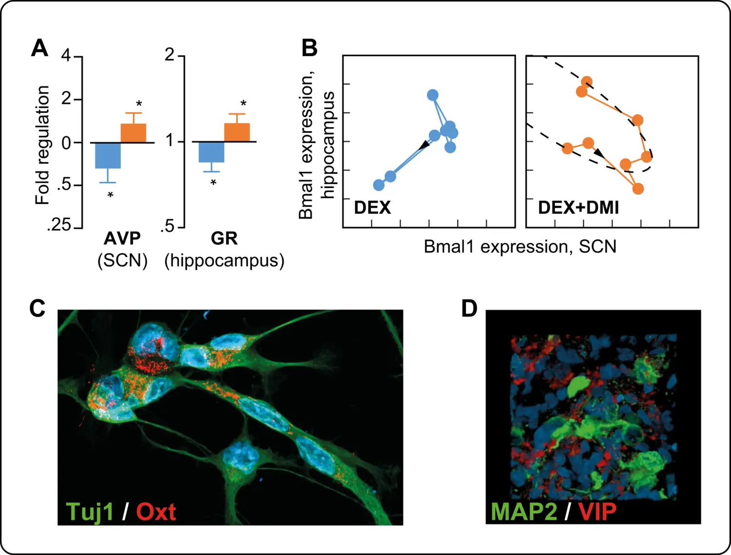
The onset of depression is preceded by long-lasting alterations in circadian patterns of activity and weaker coupling between the central clock (located in the hypothalamic suprachiasmatic nucleus) and peripheral oscillators. Collaborative clinical investigations show abnormal circadian activity patterns in depressed patients as well.

Publications

Selected publications

Sex Differences in Long-term Outcome of Prenatal Exposure to Excess Glucocorticoids-Implications for Development of Psychiatric Disorders.
Elberling F, Spulber S, Bose R, Keung HY, Ahola V, Zheng Z, Ceccatelli S
Mol Neurobiol 2023 Dec;60(12):7346-7361

Patterns of activity correlate with symptom severity in major depressive disorder patients.
Spulber S, Elberling F, Svensson J, Tiger M, Ceccatelli S, Lundberg J
Transl Psychiatry 2022 Jun;12(1):226