Division of Radiology

The Division of Radiology combines research and education with daily clinical care. The division was founded in 2017 and works in close collaboration with Karolinska University Hospital in Huddinge, where it is integrated into the hospital's operations. This integration provides access to patients, image data and infrastructure, offering excellent opportunities for research, teaching and learning.

At the Division of Radiology, experts from radiology, nuclear medicine and other clinical areas work together to develop and improve healthcare. The division's faculty council consists of professors, subject representatives and researchers from Karolinska Institutet, including adjunct and visiting professors.

Education and research are a natural part of the daily work at the clinic. Multidisciplinary rounds, where different specialists meet, create an arena for collaboration between researchers, doctoral students, hospital staff, universities and companies. Together, we work to develop new solutions, evaluate them and make improvements available to patients in regular healthcare.

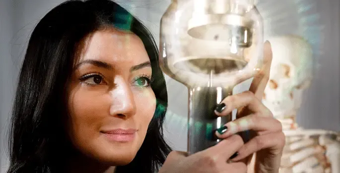
Collaboration for the future of medical imaging

Together with SMAILE (Stockholm Medical Artificial Intelligence and Learning Environments), we offer image material and courses for researchers from different backgrounds who are interested in medical image analysis.

Through our involvement in Jonasson's imaging centre at KTH Campus South, we have access to more researchers from different disciplines and advanced equipment for both clinical and preclinical imaging research. This strengthens our work in developing new methods and solutions in medical imaging technology.

Research Group Function and Technology

Since 2023, the Research Group Function and Technology has been part of our division. The group consists of researchers from Karolinska Institutet with expertise in clinical physiology, nuclear medicine, hospital physics and medical technology.

In addition to research, the group is also responsible for teaching at Karolinska Institutet, the Royal Institute of Technology (KTH) and Stockholm University (SU). Through their broad collaboration, they contribute to the development of future medical technology and education.

Organisation

Profile image

Torkel Brismar

Professor/Senior Physician

Address

Karolinska Institutet
Department of Clinical Science, Intervention and Technology (CLINTEC)
Division of Radiology
Karolinska University Hospital. C1:46, Huddinge
S-141 86 Stockholm

Publications

All publications from group members

Staff and contact

Head of Division

All members of the group

Research

Our research in medical radiology and nuclear medicine involves all modern imaging modalities with a focus on high-tech equipment, such as MRI, dual-energy high-resolution CT, SPECT-CT, PET-CT, and extremity cone-beam CT.

Upper gastrointestinal radiology research focuses on tumour detection and treatment, but is also strong in the field of transplantation.

Liver function and its regeneration after surgery, detection of liver and pancreatic cancer, primary sclerosing cholangitis, treatment effects and prediction in oesophageal cancer are included. In interventional radiology, translational chemoembolisation (TACE) therapy is performed on liver and kidney tumours in clinical trials. Additional clinical and preclinical research areas include cardiovascular and thoracic imaging, skeletal and trauma, PET detection of lung tumours, and new imaging and analysis methods implemented in collaboration with KTH.

Abdominal Radiology

In abdominal radiology, there are multiple ongoing projects to develop new CT and MRI-methods to quantify organ function as well as to diagnose cancer earlier. Also, our aim is to improve staging and evaluation of tumor response to treatment.

Our research profile is in line with the profile of our hospital, i.e. transplantation and upper abdominal surgery. Principles of the so-called ”precision medicine” (i.e., the right patient is provided with the right treatment at the right time) play a central role. Research group leaders are associate professor Torkel Brismar and Nikolaos Kartalis, M.D., Ph.D. Below are some examples of the ongoing projects in the Division of Radiology at CLINTEC:

Perfusion CT

CT of a solid tumor in the liver or pancreas with very high temporal resolution after the injection of iodine-based contrast media, we try to improve characterization and/or predict/assess earlier whether the tumor will respond to therapy or not.

Optimization of contrast media dose at CT

In order to improve visualization of organs and their diseases, we administer intravenously iodine-based contrast agents. In these projects, we try to optimize the amount of contrast agent to patient size/ composition and heart function in order to inject a sufficient amount of contrast agent at an appropriate rate for the specific clinical setting.

Esophageal and colon cancer

CT-based analysis of tumors’ morphometric structure may improve our ability to identify which patients have aggressive tumors and who will have a good response to treatment. This can potentially save patients that have responded well to non-surgical treatment from surgery (regarding esophageal cancer) and/or lead to a timely change to other forms of treatment in those patients whose tumors did not respond to standard treatment.

Pancreatic cancer

CT and MRI are used to improve detection, staging and characterization of solid and cystic pancreatic tumors. Furthermore, we investigate pancreatic function by injecting a substance similar to a body hormone (namely, secretin) and then quantifying the excretion of pancreatic fluid on MRI images.

Primary sclerosing cholangitis (PSC)

In patients with PSC, there is a chronic inflammation of the bile ducts. The inflammatory changes lead to a progressive destruction of parts of the liver and predispose to cholangiocarcinoma. By applying advanced post-processing of MR images, we try to predict which patients have a high risk to develop a clinically significant liver function deterioration or cholangiocarcinoma.

Liver function

There are many diseases that lead liver function deterioration, such as inflammation (hepatitis), diabetes mellitus, obesity and alcoholism. Chemotherapy may also lead to liver dysfunction. In order to assess treatment effect and estimate the proportion of liver that needs to remain within the body after liver surgery, it is important to calculate the degree of fatty infiltration, fibrosis and function of the liver. We try to quantify all these parameters by analyzing how the MRI signals of fat and water interact with each other, how fast a shake-wave propagates through the liver parenchyma (the so called “MR elastography”) and by calculating the excretion of hepatobiliary-specific contrast agents.

Diagnostic and interventional oncologic radiology (DIOR)

Primary tumors and metastases may be treated microinvasively through heating (thermal ablation), freezing (cryoablation) and direct alcohol injection. Proper positioning of the needle/probe is performed under ultrasound or CT guidance. Some tumors maybe difficult to identify either because they are too small and/or they are only visible during a particular phase at dynamic contrast-series, or at MRI. In this project, we fuse images from CT or MRI with ultrasound. Thus, advantages of both techniques can be combined, such as the high spatial resolution of CT/MRI with the real-time imaging of ultrasound. In order to achieve needle/probe placement in very small tumors, minimizing respiratory-related motion is necessary. For that, patients may have a general anesthesia and receive high-frequency jet ventilation.

Primary investigators are Torkel Prismar and Nikolaos Kartalis.

Musculoskeletal Radiology

Musculoskeletal research is mainly is focused on studies on osteoporosis and body composition and is lead by Torkel Brismar.

Nuclear Medicine

In nuclear medicine, we have a wide cooperation in different fields: infectious diseases, orthopedics, gastro surgeons, urologists, transplant surgeons, stem cell researchers, and neurologists. Much of the research is devoted to using hybrid imaging with SPECT / CT and PET / CT for early diagnosis, staging, and monitoring the effect of treatment in oncology as well as neuroscience in geriatrics and orthopedics / infection. In the current situation there are six Ph. D students in these field; and we have two PhD doctor, and a registered Ph. D student employed in this section. Research leader is Professor Rimma Axelsson

Below is a selection of projects

Hybrid imaging SPECT/CT

  • MIDOR – Molecular imaging in Differentiation of Oncocytoma from Renal Cell carcinoma

Hybrid imaging with PET/CT

  • Evaluation of treatment effects with proliferation marker 18F-FLT in patients with esophageal carcinoma.
  • Natural History of healing after Prosthetic surgery followed by 18 F-FDG-WBC PET / CT
    o infections in orthopedic prostheses - comparative study of 18 F-FDG-WBC with SPECT / CT and 99mTc-HMPAO
  • WBC with SPECT / CT
  • 68Ga-PSMA-11 PET/CT in high risk patients with primary prostate cancer
  • 68Ga-PSMA-11 PET/CT in patients with biochemical relapse of prostate cancer

Thorax

Regarding the cardiovascular area, we have a close cooperation with cardiologists and a great deal of research has been devoted to establishing and developing the method of CT of the coronary arteries (CTA) with ECG-triggered technology. This led in 2011 to the thesis ”Computed tomography of the coronary arteries ” by Jonaz Ripsweden.

Today, research is partly focused on CTA in the study SMINC 2, which is collaboration between 5 Stockholm hospitals. We examine the coronary arteries of all MINOCA patients in Stockholm who have suffered a heart attack but have no angiographic stenoses (Myocardial Infarction with Non-Obstuctive coronary angiography). The goal is to find out what this state consists of and how to prevent it. The radiological part of SMINC 1 led in 2015 to the thesis "Coronary computed tomography in patient with myocardial infarction and non-obstructed coronary arteries" by Elin Bacsovics.

Research in contrast optimization in CTA led in 2015 to the thesis "Intravenous contrast media optimization at computed tomography" by Anders Svensson.

We have research collaboration with cardiothoracic surgeons and cardiologists regarding time-resolved studies (so called 4D technology) of percutaneously and surgically implanted biological aortic valves to study thrombosis and reduced mobility of the leaflets in the postoperative course.

Research leader is Docent Kerstin Cederlund.

Since April 2015 we have carried out CT scans of coronary arteries, lungs and body composition in 5000 subjects between 50 and 65 years in the national study SCAPIS. This is done in the radiology department at Karolinska University Hospital, Huddinge.

Education

We provide teaching in medical imaging and functional medicine at undergraduate (bachelor's and master's), specialist and postgraduate levels. We are responsible for undergraduate education in medical imaging and functional medicine (radiology) and also participates in other educational programmes at KI and KTH's School of Technology and Health (STH) for doctors and nurses (especially radiographers).

The Division of Radiology is very active in teaching medical students the pros and cons of different imaging modalities. Our teaching is both interactive with continuous feedback that enables us to improve our pedagogic methods to ensure the best possible results in learning.

The teaching takes place mainly on fourth and seventh academic terms.