Beta Cell in-vivo Imaging/ Extracellular Flux Analysis (Seahorse)
These two unique facilities to support research in the area of diabetes and metabolism are located at the Rolf Luft Center, Department of Molecular Medicine and Surgery in close vicinity of the new Karolinska Hospital at Solna.
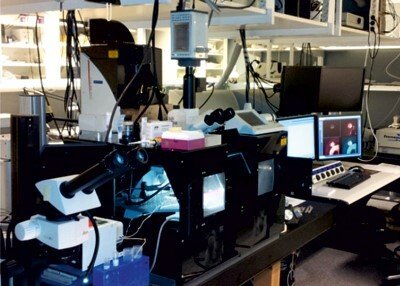
Beta Cell in-vivo Imaging
This facility with state-of-the art microscope technology for visualizing beta cells in different experimental settings is supported by the programme. The facility could also be used for imaging other cell-types. Instrumentation for in vitro fluorescence-based microscopy includes one confocal microscope, one TIRF (total internal reflection fluorescence) and two microscopes for dynamic microfluorimetry. High-through put imaging can be performed using high content screening (BD Pathway 855 instrument) as well as fluorescence activated cell sorting (FACS with 5 lasers and 16 detection channels) for analyses and sorting of cells. Two upright microscopes are equipped for in vivo imaging using confocal and multiphoton laser-scanning microscopy. A unique platform has been developed at the facility to enable non-invasive studies of islets transplanted to the anterior chamber of the eye (ACE, mouse or rat) where the cornea serves as a natural ‘body-window’ for in vivo imaging.
Contact imaging facility:
Per Olof Berggren
Professor, SeniorMartin Köhler
Research SpecialistMeasuring Metabolism with SeaHorse Flux Analyzer
Understanding cell metabolism is essential for understanding diseases such as diabetes, obesity, cancer, Parkinson’s and Alzheimer’s, just to mention a few. Mitochondria are a key organelle responsible for cell energy production. They are important in aging processes, cellular death/apoptosis and the demarcation of tissue phenotypes. Using mitochondria or mitochondria plus metabolism as PUBMED filters, one can notice an increase of almost 300% in publications in the last 20 years. This highlights the importance and development of this field and the necessity to continually have updated tools at hand. Mitochondrial respiration can be measured using a novel and powerful technique, namely state-of-the-art extracellular flux analysis. This allows monitoring in real time of O2 consumption rate (OCR) which, in the presence of a given substrate, is a reflection of the efficiency of utilization of that substrate by mitochondria. Mitochondrial indexes such as the fraction of respiration coupled to ATP production or the maximal respiratory capacity can be determined through select pharmacological manipulations. In addition, this technique allows, under certain conditions, estimation of the glycolytic flux (ECAR) and thus the ratio between respiratory capacity and glycolytic flux and hence the comparison between the pathways of cellular energy production. Importantly, this technique can be implemented in a 96-well format, thus enabling the possibility to carry out high-throughput screening studies, and with spheroid plates, enabling single or multiple spheroids or pancreatic islet measurements. The instrument in both formats (24 and 96 wells) is now part of the Beta Cell in-vivo Imaging/ Extracellular Flux Analysis core facility.
Agilent is the provider of the Seahorse Flux Analysers and we serve as a reference laboratory for Karolinska Institutet.
User agreement form, Costs and Regulations: see the documents below.
For service improvement, please fill in the feedback form below and send by email or anonymously print it and leave it in our mailbox.