Gel documentation and Western imaging core facility at BioClinicum
The OnkPat/Theme Cancer core facilities are available for staff at the Department of Oncology-Pathology in addition to research Theme Cancer in BioClinicum. New users need an introduction to get authorized. Contact information to responsible persons can be found below. Before using this core facility send your certificate from KI laboratory safety introduction to Paula Mannström. Access to the building is also required.
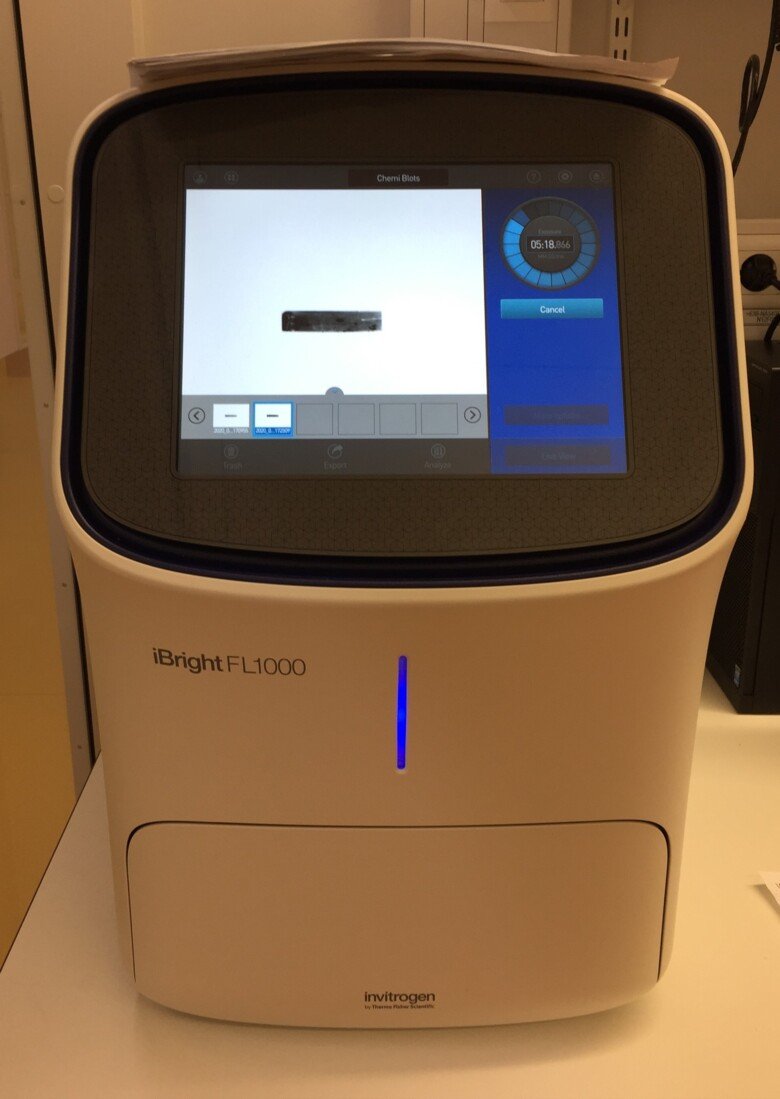
iBright FL1000/ Invitrogen by ThermoFisher Scientific
Room nr: U230 06 3700, BioClinicum J6:30
Responsible person: Susanne Öhlin
Booking calendar: Clustermarket
General information
Equipment from Invitrogen allows both gel documentation and Western blot imaging. This is a high-performance instrument for capturing fluorescent and luminescent images and analyzing data from gels and blots. Equipped with 16-bit CCD camera and Long-life green LED-based trans-illuminator. Applications: colorimetric staining of gels, SYBR dye staining of gels, Chemiluminescence and fluorescence detection (RGB, a visible range; near-IR fluorophores, such as Alexa Fluor and Alexa Fluor Plus dyes and DyLight dyes) for Western blot imaging. Cloud connectivity with Invitrogen iBright Analysis Software enables export and storage of data, as well as the ability to access, review, analyze, and share data through the web-based Thermo-Fisher Cloud platform.
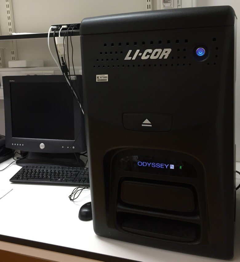
Odyssey Fc/LI-COR Biosciences
Room nr: U230 06 3700, BioClinicum J6:30
Responsible person: Xiaohao Wang
Booking calendar: at the location
General information
The Odyssey imaging system allows for both the enhanced chemiluminescent (ECL) and the near-infrared (NIR) fluorescence detection using two-color detection methods for Western blots. The patented optics of the Odyssey Fc deliver uniform low background images. For Western blotting, apart from ECL, one can multiplex to detect two different protein targets in each sample lane by using secondary antibodies labeled with spectrally-distinct NIR fluorescent dyes. The NIR fluorescence delivers consistent signals that aren’t affected by timing. The ImageStudio software is installed on a MacOS computer connected to the Imaging system. The software allows quick and easy acquisition, adjustment and organization of images. ImageStudio versions can be downloaded from the LI-COR website and installed on both PC and MacOS users’ computers.
iBright FL1500/Invitrogen by ThermoFisher Scientific
Room nr: U220 06 3700, BioClinicum J6:20
Responsible person: Wen Zhong
Booking calendar: Clustermarket
General Information
The iBright FL1500 Imaging System supports the main imaging applications of fluorescent, chemiluminescent, and colorimetric western blots, in addition to fluorescent stained nucleic acid gels, fluorescent stained protein gels, colorimetric stained protein gels, and colorimetric membrane stains.
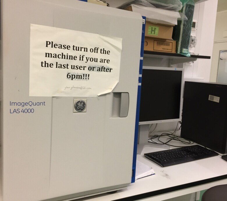
ImageQuant LAS-4000/GE HealthCare
Room nr: U220 06 3300, BioClinicum J6:20
Responsible person: vacant
Booking calendar: at location
General information
ImageQuant LAS 4000 is a digital imaging system for sensitive, quantitative imaging of gels and blots, by fluorescence and chemiluminescence and white epi-illumination imaging applications. It allows capturing and precisely quantitating weak and strong signals with high resolution and sensitivity and low noise. User-friendly image capture software ImageQuant LAS 4000 performs several capture modes for achieving optimal sensitivity and dynamic range including increment, repetition, and program modes.
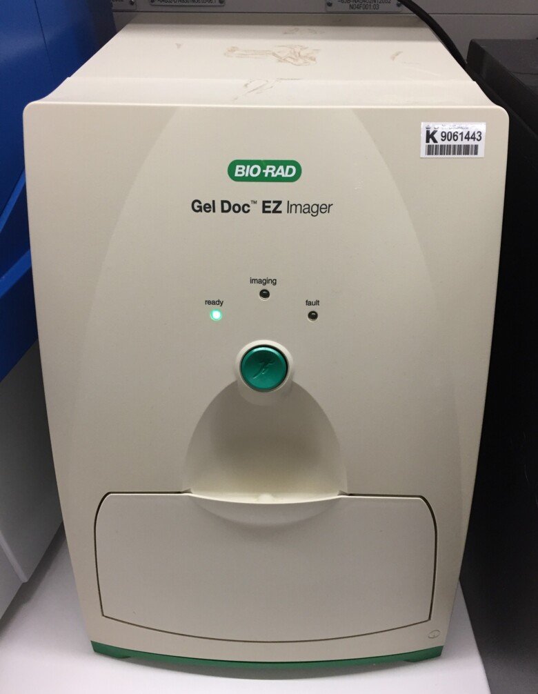
Gel Doc EZ imager/BioRad
Room nr: U220 06 3306, BioClinicum J6:20
Responsible person: vacant
Booking calendar: at location
General information
This GelDoc Imager is a compact and automated imaging system for obtaining images of Nucleic acid-gels or protein gels and a possibility for analysis. The system provides four application-specific trays, which include a UV tray for staining of DNA gels and fluorescence imaging, a white tray for Coomassie, copper, silver, and zinc stains, a blue tray for nucleic acid applications that use SYBR® stains, and a stain-free tray for stain-free imaging. The Stain Free Sample Tray is a useful tool for visualization of protein bands on gels and blots. TGX Stain-Free Precast gels from Bio-Rad can be activated in the Gel Doc EZ after electrophoretic separation of proteins is completed. Once activated, the same protein bands can be visualized on protein membrane after transfer (Nitrocellulose or PVDF). Thus, the stain- free technology enables one to monitor electrophoretic separation and transfer efficiency without any additional staining. The ImageLab™ software makes imaging and analysis pretty easy with such options as auto image capture, auto analysis or user preferences. ImageLab software is free and can be downloaded on your PC or Mac for analysis purposes from Bio-Rad.com.