Microscopy core facility in BioClinicum
This lab is a core facility for OnkPat and Theme Cancer in BioClinicum. The lab is accessible for authorized persons to work with light (bright field, phase contrast), epifluorescence and confocal microscopy. New users should contact one of the responsible persons to become an authorized user and to be added to the booking calendar.
Room nr: U230 06 2100
Booking calendar: Clustermarket
Work procedures: There are defined rules and work procedures for starting the microscopes, operating them and using the software as well as finishing the work, all of which have to be strictly followed.
Equipment
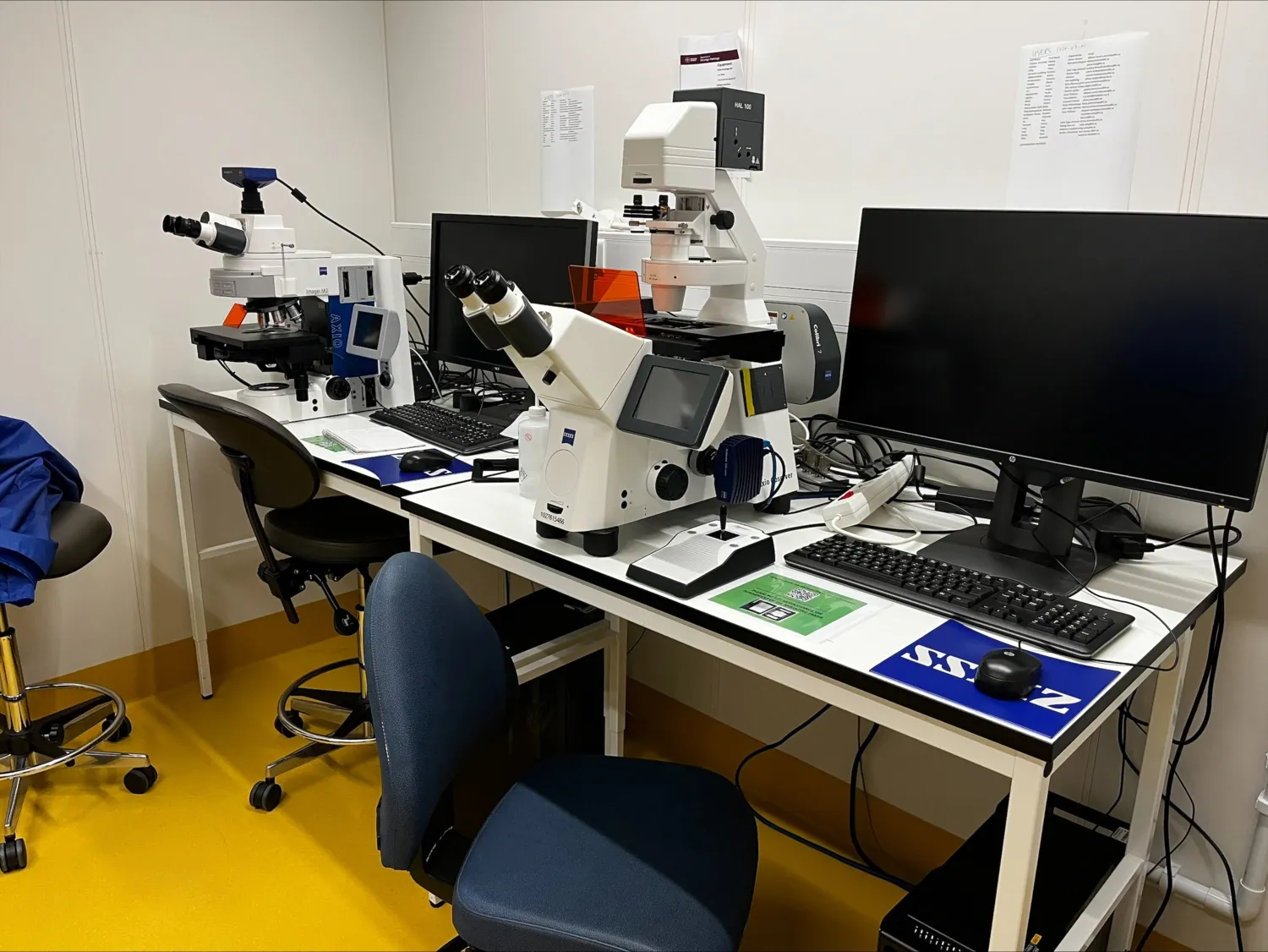
Zeiss AxioImager M2
- for slides
- monochrome camera (IF)
Software functions
- image quantification
- deconvolution
Fluorescence filters: DAPI, GFP, Rhodamine, FITC, Texas Red, Alexa Fluor 568
Responsible persons: Lena Lennartsson and Paula Mannström
Zeiss AxioObserver Z1 (inverted)
- for plates AND slides (change mounting frames yourself)
- monochrome camera (IF including far red).
- color camera (BF)
- LED illumination/Colibri 7
- optical sectioning/ApoTome
- phase contrast
Responsible persons: Lena Lennartsson and Paula Mannström
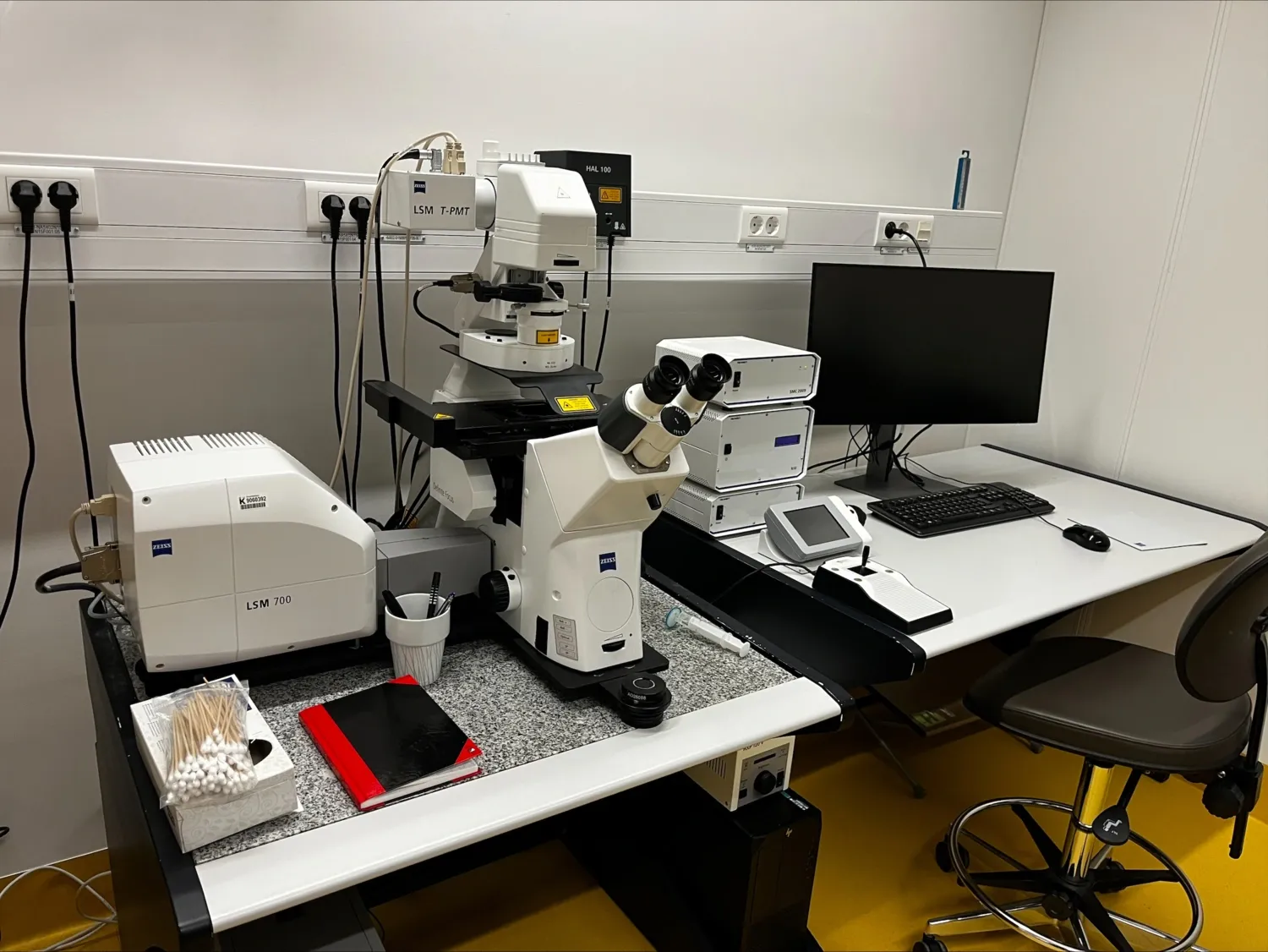
Zeiss LSM700 Confocal Microscope
The LSM700 confocal microscope can acquire automatic tile/mosaic images of dyes/fluorochromes within the full visible range (blue to far-red). It can also perform spectral separation to separate fluorochromes with overlapping spectra.
Specifications
- Stage inserts for: slides and multiwell plates.
- Lasers: 405, 488, 555, 639 nm.
- Fluorescent channels: from blue (DAPI), Green (GFP), orange-red (Cy3) to far-red (Cy5).
- Objectives: 10x Air, 20x Air, 40x Water and 63x Water.
- Acquisition Software: Zen Black
Special Features
- Motorized scanning stage that accommodates inserts for slides or multiwelll plates.
- Possibility of collecting Zstacks, tiles, timelapse and positions.
- Lambda mode/Spectral Imaging, useful for applications such as FRET.
Responsible persons: Ophelie Le Chapelain, Francesca Del Gaudio and Pedro Fonseca