Widefield Microscopes at BIC
The BIC facility has a diverse range of widefield microscopes for fast fluorescence and brightfield imaging of fixed and live imaging of biological samples.
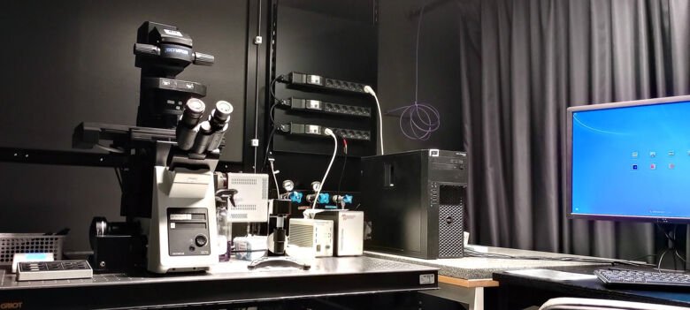
Olympus IX73
The Olympus IX73 widefield inverted microscope can take both colorimetric and fluorescence images from blue to far red dyes/fluorochromes. It can acquire multi-position and/or tile images and is suitable for slides, dishes or multiwell plates.
Specifications
- Stage inserts for: slides (up to 3)/multiwell plates/35-60mm dishes
- Light Source: CoolLED pE-300 White (365, 460, 525, 660nm)
- Fluorescent channels: DAPI, FITC, TRITC and Cy5
- Transmitted channels: brightfield, phase contrast
- Objectives: 2x, 4x with phase contrast, 10x with phase contrast, 20x, 20x with phase contrast, 40x with phase contrast (all Dry/Air)
- Camera: Olympus XC-10 IR (14 bit, 1376x1032, 6.45 mm pixels)
- Acquisition Software: Olympus CellSens
- Special Features:
- Supports colorimetric image acquisition
- 20x extra-long working distance objective (WD up to 7.8mm)
- Allows automatic collection of tiles and positions
Leica DMI6000
The Leica DMI6000 widefield fluorescence system allows fluorescence and transmitted-light imaging of slides and multi-well plates of fixed and live samples. Light-blocking climate chamber supports live imaging for a few hours to multiple days. The system is equipped with temperature control unit but requires CO2-independent medium since the setup has no CO2 supply.
Specifications
- Stage inserts for: slides/multiwell plates/35-60mm dishes
- Light Source: Leica LEDs: 365, 435, 470, 505 & 530nm; Leica halogen lamp EL6000
- Fluorescent channels: DAPI, CFP, FITC, YFP, TRITC and Cy5
- Transmitted channels: brightfield, DIC
- Objectives: 5x, 10x, 20x, 40x (all Dry/Air)
- Camera: Roper Evolve 512 (512x512, 16 mm pixels)
- Acquisition Software: Leica LAS X 3.6
- Special Features:
- Black climate chamber with temperature and humidity control for live cell imaging
- 20x extra-long working distance objective (WD=6.9mm)
- Advanced tiles & positions (including focus maps and stitching)
- Filter set for CFP/YFP detection
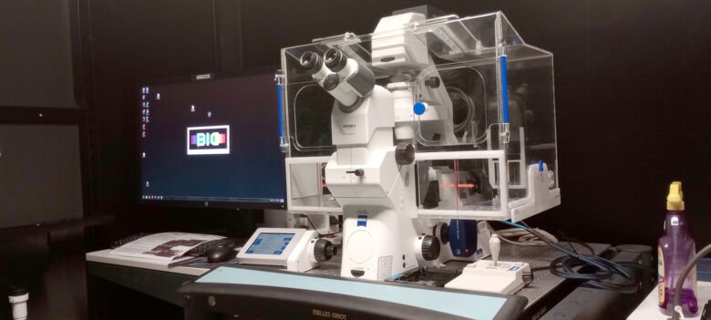
Zeiss Cell Observer
The Zeiss Cell Observer is a widefield inverted microscope equipped with temperature, humidity and CO2 control for live imaging of cells and spheroids/organoids on slides and multi-well plates. This system is equipped with extra-long working distance objectives and facilitates bright field, epifluorescence, DIC and phase contrast microscopy.
Specifications
- Stage inserts for: slides/multi-well plates/35-60mm dishes
- Source: Colibri 5 LED (385, 469, 555, 631nm)
- Fluorescent channels: DAPI, CFP, FITC, YFP, TRITC, mCherry and Cy5
- Transmitted channels: brightfield, DIC, phase contrast
- Objectives: 2.5x, 10x with phase contrast, 20x, 20x with phase contrast, 40x, 40x with phase contrast (all Dry/Air)
- Camera: Zeiss AxioCam 702 (14-bit, 1920 x1216, 5.86 mm pixels)
- Acquisition Software: Zeiss Zen Blue v2.3
- Special Features:
- Transparent climate chamber with temperature, humidity and CO2 control for live imaging
- Extra-long working distance objectives (20X: WD=7.9mm; 40X WD=2.9mm)
- Advanced tiles & positions (including focus maps and stitching)
- Filter set for CFP/YFP detection
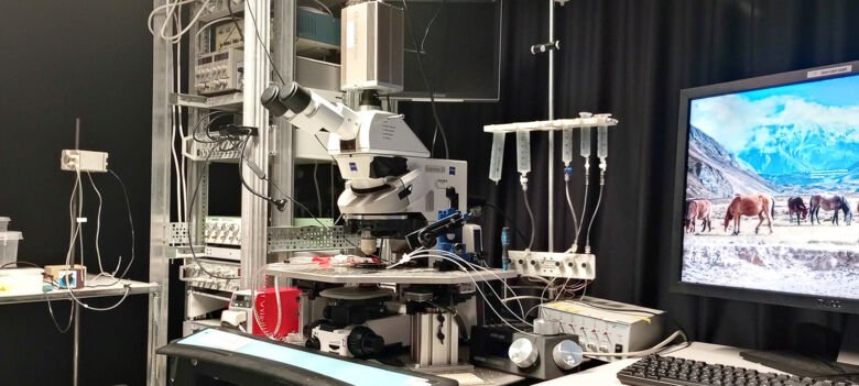
Zeiss AxioExaminer.D1 (Calcium Upright)
The “Calcium upright” microscope is an upright manual fluorescence microscope equipped for imaging of very rapid fluorescence events, such as imaging of calcium fluctuations. Equipped with a heated 35mm petri dish holder and can also image multiwell plates, slides and petri dish <10cm without heating.
Specifications
- Stage inserts for: slides/multiwell plates/35-100mm dishes (heated for 35mm dish)
- Light Source: Sutter Instruments DG4/17 xenon arc lamp
- Fluorescent channels: Fura2 (blue), Fluo3/4 (green), Fura2/Fluo3/4 (blue/green), Fura2/Rhod2 (blue/red), Fluo3/4 /Rhod2(green/red). Extra: CFP.
- Transmitted channels: bright field, IR-DIC
- Objectives: 20x water dipping, 63x water dipping (can be exchanged with other dipping objectives)
- Camera: Photometrics Quant-EM 512 SC (512x512, 16x16µm pixel size)
- Acquisition Software: MetaFluor 64bit v7-10-1
- Special Features:
- A special illumination lamp supports imaging of very fast events
- Equipped with heated 35mm Petri dish holder
- Infra-red DIC to minimize phototoxicity and improve the contrast
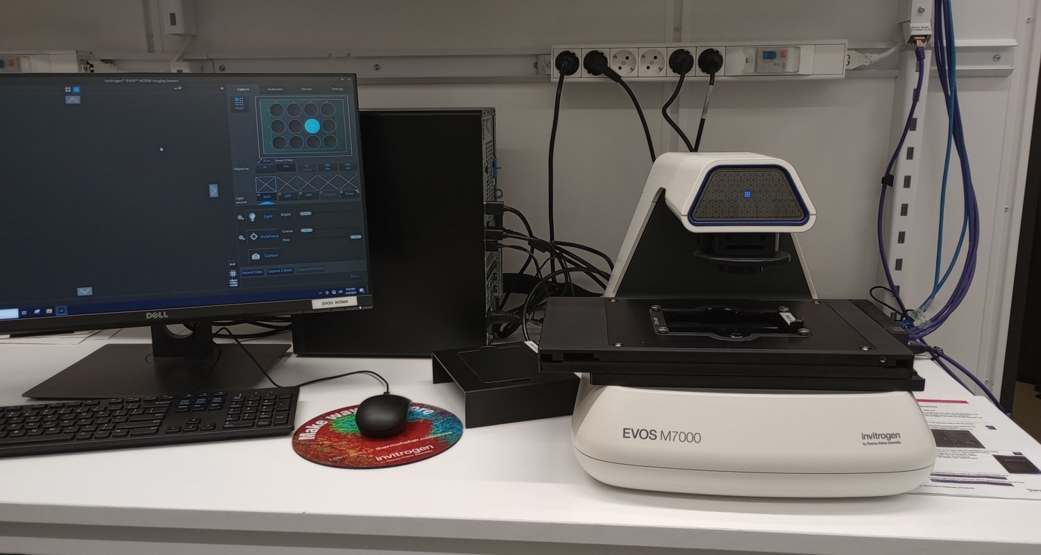
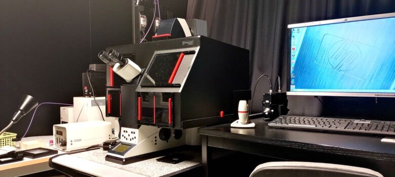
ThermoFisher EVOS M7000
The EVOS M7000 is a widefield inverted imaging system equipped with long-distance objectives and motorized stage. It has both a color camera and a monochrome camera, supporting blue, green, and red fluorescence, brightfield, color brightfield, and phase contrast imaging modes. The system comes with a variety of stage inserts suitable for nearly all types of slides, plates, flasks, and dishes. The EVOS M700 is specially intended for quick sample assessment by BIC users and is available free of charge.
Specifications
- Stage inserts for: slides/multi-well plates/35-60-90mm dishes/flasks
- Light Source: LED cubes (DAPI, GFP, RFP, Cy5)
- Fluorescent channels: DAPI, GFP, RFP, Cy5
- Transmitted channels: brightfield, color brightfield, phase contrast
- Objectives: 4x/0.13, 10x/0.3, 20x/0.6, 40x/ 0.6 (all Dry/Air)
- Acquisition Software: EVOS M7000
- Special Features:
- Wide selection of the vessel templates available in the software
- Color brightfield imaging mode
- Supports single color, multi-color, area scan with montage or tile-stitch, time lapse, Z-stacking and movie capture
- Extra-long working distance objectives (20X: WD=6.6mm; 40X WD=2.7mm)