Image Processing and Analysis at BIC
As a BIC user, you have access to workstations equipped with diverse image analysis software, including both licensed and free-ware products. These tools support image processing, intensity measurements, object counting, and the creation of movies and 3D models. The workstations are conveniently situated next to the microscopes at BIC2.
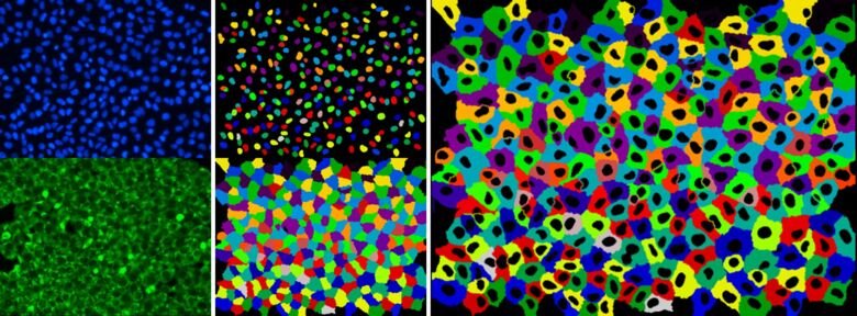
Image analysis tools
All workstations are equipped with the vendor’s ‘lite’ versions of the acquisition software (Zeiss Zen Lite, Olympus Micro, LAS X Lite, NIS Element Viewer, IncuCyte), along with the basic analysis tools (ImageJ/Fiji, MATLAB, CellProfiler, and QuPath). Additionally, licensed software such as Imaris, Amira, AutoQuant, and NisElements is available. If you require specialized software for your analysis, it can be installed upon request.
Workstations at the BIC facility
All workstations are free to use for BIC users but need a reservation in iLab. The working stations can be accessed remotely via the VNC Viewer, ask the BIC facility staff for the access instructions. OBS! Do not turn workstations off when finished, only log out of the BIC user account.
Workstation 3 (Imaris with Clearview deconvolution)
WS3 features Imaris, MATLAB, viewer versions of ZEN and LAS microscope software, Fiji, CellProfiler, QuPath, KNME Analytics plus other software.
RAM 256GB, Intel Xeon W-2133, 3.60GHz, 6 cores, 12 threads. Graphic card NVidia Quadro P2000, 5GiB
Workstation 4 (NIS Elements)
WS4 has NIS Elements (Nikon), FluoView (Olympus), MERSCOPE visualizer/vpt/cellpose2 (Vizgen) in addition to basic analysis tools.
RAM 512GB, Dual Intel Xeon E5-2643 v4 (3.40GHz, 12 cores, 24 threads) and NVidia Titan RTX, 24GB.
Workstation 6 (Imaris, AutoQuant deconvolution)
WS6 is a workstation with Imaris full licence, however without the built-in Clearview deconvolution. Instead, it has the AutoQuant deconvolution program, in addition to the viewer versions of the microscope software plus Fiji, CellProfiler, QuPath, KNME Analytics etc.
RAM 768 GB, Intel Xeon Gold 6250, 3.90 GHz, 8 cores, 16 threads. Graphic card NVIDIA Quadro RTX 6000, 24GiB
Workstation 7 (Imaris with Clearview deconvolution)
WS7 is a powerful analysis station with a full Imaris license including the ClearView deconvolution module. It also features MATLAB and viewer versions of the microscope system software, plus Fiji, CellProfiler, QuPath, KNME Analytics etc.
RAM 1 TB, Intel Xeon Gold 6250, 3.90 GHz, 8 cores, 16 threads. Graphic card NVIDIA RTX A6000, 48 GiB
Workstation 8 (Imaris with Clearview deconvolution)
WS8 is dedicated to the big datasets' analysis like obtained by light sheet microscopy. It has installed Imaris and programming software.
RAM 1 TB, Intel Xeon Gold 6250, 3.90 GHz, 16 cores, 32 threads. Graphic card NVIDIA RTX A6000, 48 GiB
Workstation 9 (Imaris with Clearview deconvolution, Amira)
WS9 is a powerful analysis station with features Imaris, viewer versions of the software, MATLAB, Amira, InCarta, Fragonfly plus Fiji, CellProfiler, QuPath, etc.
RAM 1 TB, Intel Xeon w7-3465X, 2.50 GHz, 28 cores, 56 threads. Graphic card NVIDIA RTX 6000 Ada, 48 GiB
BIC ImageClinic
BIC ImageClinics are regular drop-in sessions for both current BIC users and those thinking about using our services. At these sessions, you can discuss with our BIC staff various aspects of microscopy, experimental design, image analysis, presentations, and more. Welcome to drop by with your questions!
- Date: Every other Wednesday, see announcements on upcoming events and activities at BIC
- Time: 09:00-12:00
- Venue: Room C0412, Biomedicum, floor 4