High Content Automated Systems at BIC

BIC facility is equipped with a diverse collection of automated, high content imaging systems for imaging tissue sections on slides, multiwell plates (6-384 wells), and multi-day live imaging experiments.
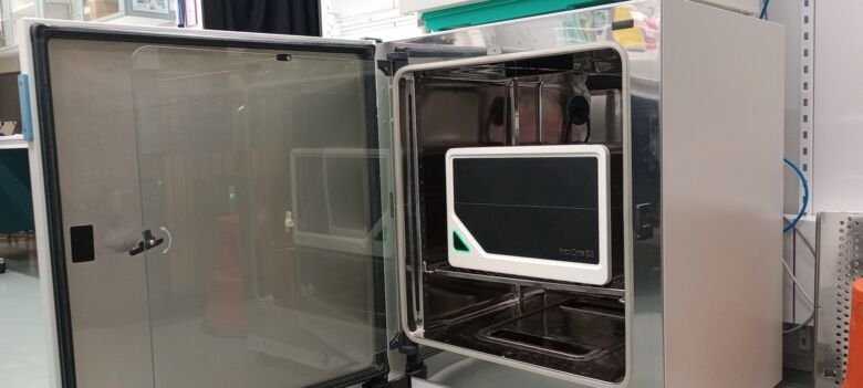
The IncuCyte S3 system is a dedicated imaging system inside a cell culture incubator with perfect temperature (37° C), 5% CO2, and humidity control., included in BIC.
Satirius IncuCyte S3. Photo: Anna Kouznetsova

Satorius IncuCyte S3

The IncuCyte S3 system is a dedicated imaging system inside a cell culture incubator with perfect temperature (37° C), 5% CO2, and humidity control. The system allows long-term transmitted light and GFP/RFP fluorescence imaging of adherent, non-adherent cells, and spheroids/organoids in diverse multi-well plate formats, ranging from 6 to 384 wells, and ultra-low attachment (ULA) U- and V-bottom plates. The system can be used by up to three different users with a maximum of 6 plates simultaneously.

Specifications

  • Stage inserts for: slides/multiwell plates (incl. 96 and 384well)
  • Light Source: LEDs
  • Fluorescent channels: GFP, RFP
  • Transmitted channels: brightfield
  • Objectives: 5x, 10x, 20x (all air/dry)
  • Acquisition & Analysis Software: IncuCyte-2019BRev2
  • Special Features:
    • located inside the cell culture incubator
    • 3 trays, 6 multiwell plate positions
    • multi-day imaging sessions for up to 3 users simultaneously
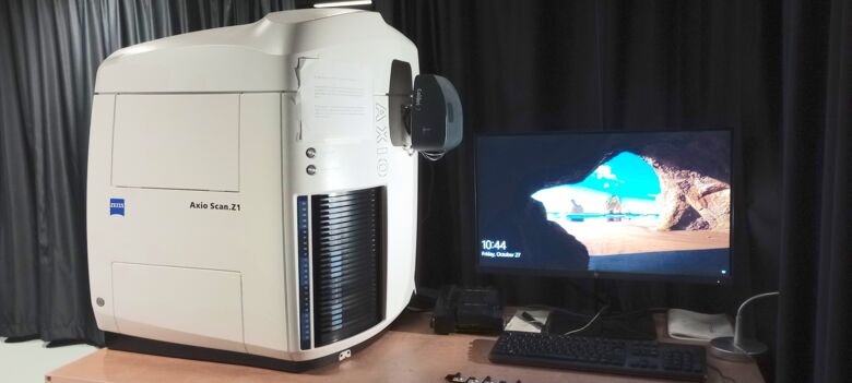
The Axio Scan Z1 is an automated slide scanner which can process up to 100 slides in a single imaging session. This instrument is capable of fluorescence imaging as well as brightfield imaging with colorimetric detection (e.g. HE/DAB) using air objectives
Ziess Axio Scan.Z1 Slide Scanner. Photo: Anna Kouznetsova

ZEISS Axio Scan.Z1 Slide Scanner

The Axio Scan Z1 is an automated slide scanner which can process up to 100 slides in a single imaging session. This instrument is capable of fluorescence imaging as well as brightfield imaging with colorimetric detection (e.g. HE/DAB) using air objectives. The system can automatically detect the location of samples on slides, create focus maps, followed by scanning the images.

Specifications

  • Stage inserts for: slides 76mm x 24-26mm
  • Light Source: VIS-LED for brightfield. Colibri 7 LED for fluorescence (385, 430, 475, 567, 630 & 735nm)
  • Fluorescent channels: DAPI, CFP, FITC, YFP, TRITC, mCherry, Cy5, Cy7
  • Transmitted channels: brightfield
  • Objectives: 5x, 10x, 20x, 40x (all air/dry)
  • Camera fluo: Hamamatsu Orca Flash 4.0 V3 (16-bit, 2048x2048, 6.5 mm pixel size)
  • Camera color: Hitachi HV-F203 (14.bit, 1600x1200)
  • Acquisition Software: Zen Blue v3.1
  • Special Features:
    • Up to 100 slides in one experiment
    • Fluorescence and colorimetric imaging
The ImageXpress Pico imaging system is a fully integrated hardware and software system for automated acquisition and analysis of fixed and live samples in diverse multiwell plate formats, ranging from 6 to 384 wells, or custom-designed plates.
Zeiss Image XpressPico. Photo: Anna Kouznetsova

Mol.Devices ImageXpress Pico

The Mol.Devices ImageXpress Pico imaging system is a fully integrated hardware and software system for automated acquisition and analysis of fixed and live samples in diverse multiwell plate formats, ranging from 6 to 384 wells, or custom-designed plates. Images can be automatically analyzed for many different parameters using pre-made application modules (multi-wavelength cell scoring, count nuclei, live/dead, translocation, mitotic index, cell cycle, granularity, neurite outgrowth etc.) and customized protocols. The system has a temperature-controlled environment, allowing time-lapse imaging of living cells in CO2-independent medium.

Specifications

  • Stage inserts for: slides/multiwell plates (incl. 96 and 384well)
  • Light Source: LEDs
  • Fluorescent channels: DAPI, FITC, TRITC and Cy5
  • Transmitted channels:
  • Objectives: 4x, 10x, 20x, 40x (all air/dry)
  • Camera:
  • Acquisition Software: Web-browser based CellReporterXpress.
  • Special Features:
    • Climate chamber with temperature control
    • Supports colorimetric image acquisition
    • Automatic image collection 

Choosing a microscope and where to find it

If you are interested in more details, we have compiled detailed information about our instruments. The information could be useful when choosing between different microscopes.

You are always welcome to contact us if you need further information or guidance among the microscopes.

Where to find our instruments.