High Content Automated Systems at BIC
BIC facility is equipped with a diverse collection of automated, high content imaging systems for imaging tissue sections on slides, multiwell plates (6-384 wells), and multi-day live imaging experiments.
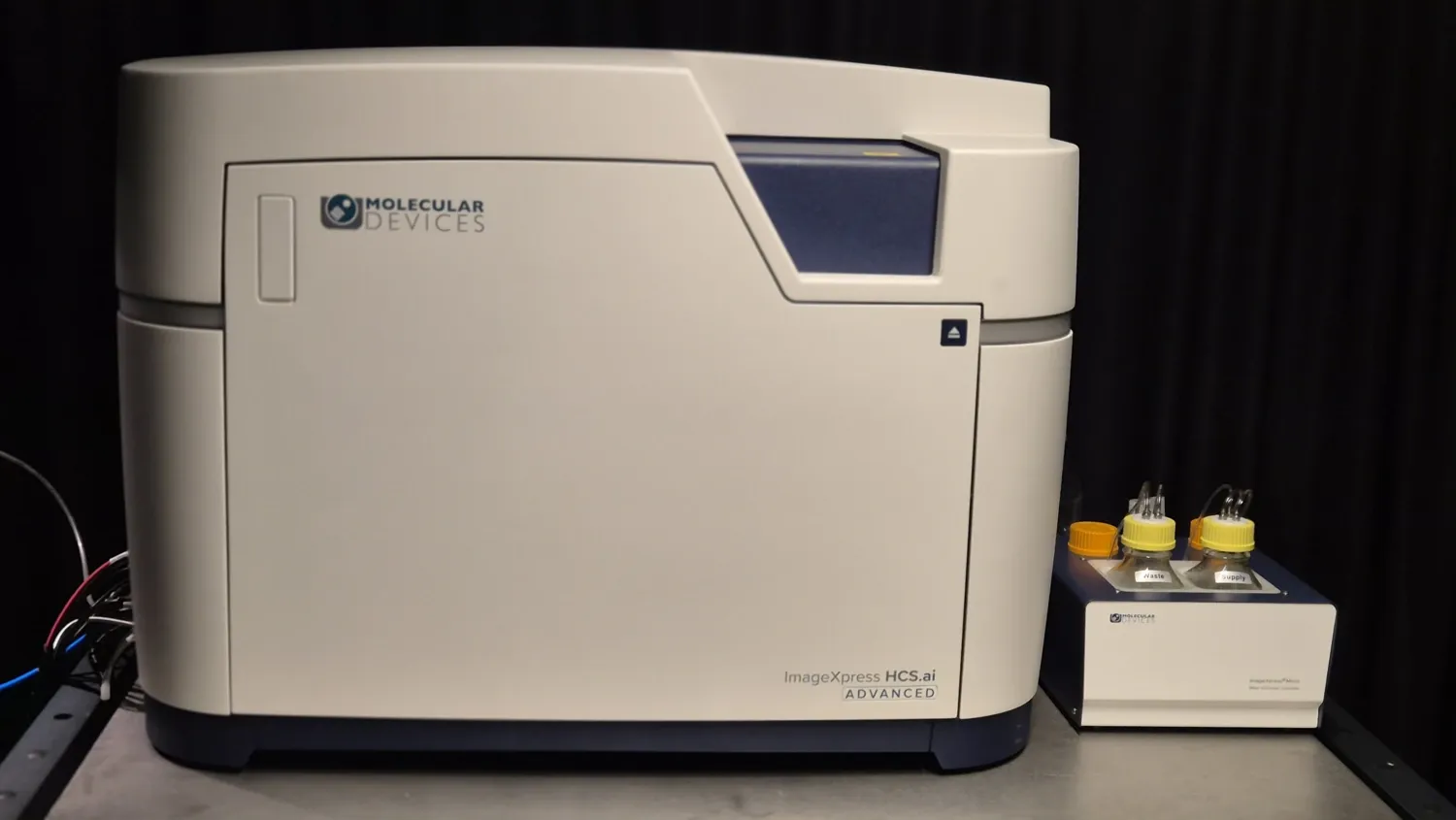
Molecular Devices ImageXpress HCS.ai
The ImageXpress HCS is an advanced high content imaging system, dedicated for fast and accurate imaging of multiwell plates (6, 12, 24, 48, 96, 384 wells) with fixed or live cells, spheroids and organoids. The system includes widefield and a two dual spinning disc confocal options for high resolution or thick samples).
The system has a robust automated hardware focusing, and high speed acquisition performance, with accurate XYZ repositioning (25nm resolution). Furthermore, it comes with an environmental control option (temperature, CO2, humidity) for live imaging.
Specifications
- Stage inserts for: multiwell plate
- Two spinning disks
- Laser: 401nm; 488nm, 514nm, 545nm, 577nm, 637nm, 748nm
- Fluorescent detection: DAPI, CFP, FITC, YFP, TRITC, TxRed, Cy5, Cy7
- Transmitted channels: brightfield
- Objectives: 4x Air, 10x Air, 20x LWD Air, 20x Water auto-immersion, 40x LWD Air, 40x Water auto-immersion
- Equipped with 1x and 1.5x magnifiers
- Acquisition & Analysis Software: MetaXpress Acquire and InCarta
- Special Features:
- climate control chamber (including CO2 control)
- 50/250 µm pinhole disk for high sensitivity in deep tissues
- 50/500 µm pinhole disk for high resolution in deep tissues
- InCarta machine learning AI software
- targeted acquisition
Download Molecular Devices ImageXpress HCS.ai start-up and shut-down procedures
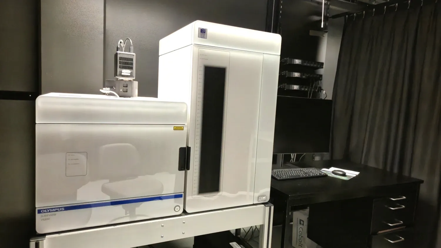
Evident SLIDEVIEW VS200 SILA
Evident SLIDEVIEW VS200 SILA is an automated slide scanner designed for high‑throughput whole‑slide imaging of histological and fluorescence samples, supporting unattended batch acquisition of up to 210 slides using the multi‑tray loader system. In addition to brightfield and widefield fluorescence imaging, the system supports darkfield and polarized light microscopy, enabling visualization of structures and features that are not readily visible with conventional transmitted‑light imaging. SLIDEVIEW VS200 performs automated sample detection, focus mapping, image acquisition, and high‑precision stitching to generate high‑resolution virtual slides. SILA (Speckle Illumination Acquisition) optical sectioning module provides confocal‑like fluorescence imaging, improving contrast and suppressing out‑of‑focus signal in thick samples.
Specifications
- Stage inserts for: slides 76x 24-26mm and 76 x51–53 mm
- Light Source: LaserLED hybrid excitation source (Excelitas X‑Cite NOVEM) with discrete excitation lines at 405, 485, 551, 584, 639, and 730 nm
- Fluorescent channels: DAPI, FITC, TRITC, mCherry, Cy5, Cy7
- Transmitted channels: brightfield, darkfield, polarized light
- Objectives: 2x (air), 4x (air) ,10x (air), 20x (air), 40x (air), 40x (oil with automatic dispenser)
- Camera fluo: 1‑inch CMOS mono camera (16‑bit, 2048 × 2048 px, 3.45 µm pixel size)
- Camera color: 2/3″ CMOS color camera (12‑bit, 2048 × 2048 px, 3.45 µm pixel size)
- Acquisition Software: VS200 acquisition
- Special Features:
- Unattended batch scanning of up to 210 slides (35 trays with 6 slides)
- Integrated barcode reading and/or slide label imaging
- Fast whole-slide acquisition (~ 80 sec for 15x15mm area at 20x in BF, excluding focusing)
- Supports brightfield, fluorescence, darkfield, and polarized light imaging
- SILA optical sectioning for high‑contrast, confocal‑like fluorescence imaging of thick samples
- Uniform fluorescence illumination using fly‑eye optics
- Automatic sample detection, automated focus mapping, and high‑precision stitching
- 40x/1.4 objective with automated oil immersion for high‑magnification batch scanning
Download Evident SLIDEVIEW VS200 SILA start-up and shut-down procedures
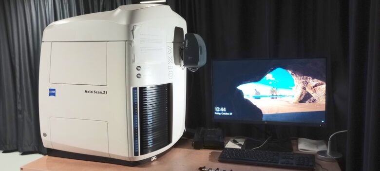
Zeiss Axio Scan.Z1 Slide Scanner
The AxioScan.Z1 is an automated slide scanner which can process up to 100 slides in a single imaging session. This instrument is capable of fluorescence imaging as well as brightfield imaging with colorimetric detection (e.g. HE/DAB) using air objectives. The system can automatically detect the location of samples on slides, create focus maps, followed by scanning the images.
Specifications
- Stage inserts for: slides 76mm x 24-26mm
- Light Source: VIS-LED for brightfield. Colibri 7 LED for fluorescence (385, 430, 475, 567, 630 & 735nm)
- Fluorescent channels: DAPI, CFP, FITC, YFP, TRITC, mCherry, Cy5, Cy7
- Transmitted channels: brightfield
- Objectives: 5x, 10x, 20x, 40x (all air/dry)
- Camera fluo: Hamamatsu Orca Flash 4.0 V3 (16-bit, 2048x2048, 6.5 um pixel size)
- Camera color: Hitachi HV-F203 (14.bit, 1600x1200)
- Acquisition Software: Zen Blue v3.1
- Special Features:
- Up to 100 slides in one experiment
- Fluorescence and colorimetric imaging
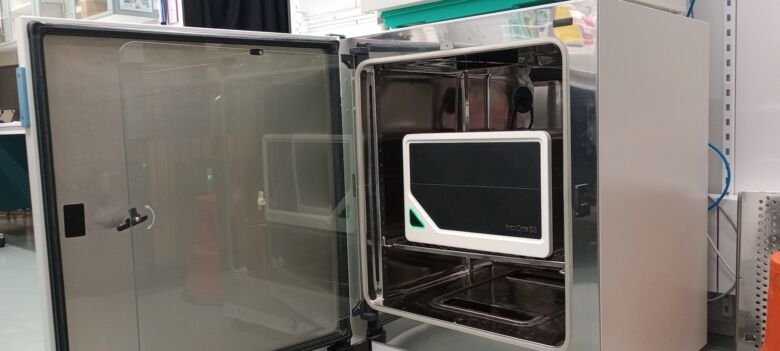
Sartorius IncuCyte S3
The IncuCyte S3 system is a dedicated imaging system inside a cell culture incubator with perfect temperature (37° C), 5% CO2, and humidity control. The system allows long-term transmitted light and GFP/RFP fluorescence imaging of adherent, non-adherent cells, and spheroids/organoids in diverse multi-well plate formats, ranging from 6 to 384 wells, and ultra-low attachment (ULA) U- and V-bottom plates. The system can be used by up to three different users with a maximum of 6 plates simultaneously.
Specifications
- Stage inserts for: slides/multiwell plates (incl. 96 and 384well)
- Light Source: LEDs
- Fluorescent channels: GFP, RFP
- Transmitted channels: brightfield
- Objectives: 5x, 10x, 20x (all air/dry)
- Acquisition & Analysis Software: IncuCyte-2019BRev2
- Special Features:
- located inside the cell culture incubator
- 3 trays, 6 multiwell plate positions
- multi-day imaging sessions for up to 3 users simultaneously