 Photo: Ulf Sirborn
Jovan P Antovic
Jovan P Antovic is research group leader of the group Clinical Chemistry and Blood Coagulation at the Department of Molecular Medicine and Surgery. Read an interview about his research.
Clinical Chemistry and Blood Coagulation conducts research mainly in the field of thrombosis and hemostasis. We are studying the usefulness of global hemostatic methods, in the diagnosis, prediction of the outcome and monitoring of the treatment in the specific clinical conditions associated with hypercoagulation.
Photo: Ulf SirbornJovan P Antovic is research group leader of the group Clinical Chemistry and Blood Coagulation at the Department of Molecular Medicine and Surgery. Read an interview about his research.
Please visit the Swedish site for more information about the research conducted within the Clinical Chemistry and Blood Coagulation research group.
Project Leader, Jovan P Antovic
Endothelial System Biology
Project Leader, Lynn Butler
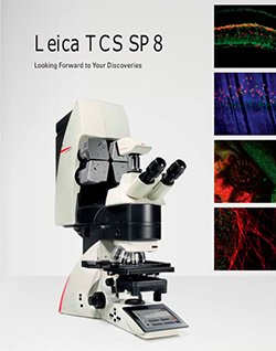
The system was designed for confocal scanning (laser scanning images) of fluorescence-marked living and fixed specimens as well as for quantitative measurements in all areas of life science. Leica TCS SP8 offers highest sensitivity with super-resolution and multiphoton or light sheet imaging. It may be used to visualise microparticles released from different cells after activation and/or apoptosis. Investigation of microparticles is of interest in different atherothrombotic diseases but also in cancer and inflammation research.
Location: BioClinicum, floor 8, Cardiovascular research group
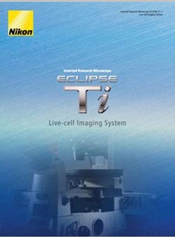
This imaging system can be used for short or long time-lapse imaging of 2D and 3D cell cultures. It is equipped with heat and CO2 controlled environmental chamber, allowing very advanced long-term automated high-throughput live imaging. Combinations of short periods fast time-lapse imaging with longer periods of slow time-lapse imaging enable the imaging of highly dynamical events in the single cell over short time and slow events over long-time periods.
Location: BioClinicum, floor 8, Cardiovascular research group
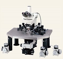
This equipment for intravital microscopy allows complex in-vivo procedures in mice and rats and imaging at the surgery site as well as in-vivo investigation of the hemostatic process (including different atherthrombosis models). This system can be easily used as core unit of in vivo vascular imaging and help many groups within KI working in the field of cardiovascular disease, thrombosis and vascular inflammation.
Location: BioMedicum
KARMITH will educate users how to best utilize services and equipment. Trained Core Facility staff will offer the necessary assistance in the formulation of scientific hypotheses, planning and execution of experiments, acquiring knowledge of methods and techniques. The KARMITH Core Facility will organise formal education in the forms of lecturing and courses.
Using of the KARMITH Core Facility will be free of charge for all users during the entire 2019. This was made possible thanks to the generous support provided by the Department of Molecular Medicine and Surgery. During 2020 the fee will be based on external fundings or by support from research groups involved.
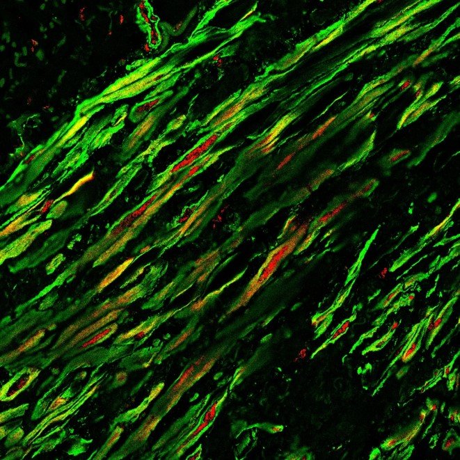
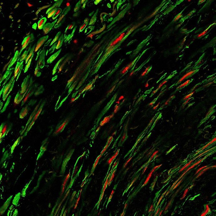
Human artery labelled with an actin marker (green) and with a protein localized in the nucleous in muscle cells (red).