Karolinska Experimental Research and Imaging Centre (KERIC)
KERIC is a core facility offering imaging analyses in experimental systems in combination with experimental surgery in large animals like swine as well as in rodents.
We offer expert services in MR, PET and CT and a fully equipped facility for experimental surgery with four full size operation units equipped with ventilators. Combination of any modality can be arranged.
Booking is done via communication with staff. See Contact
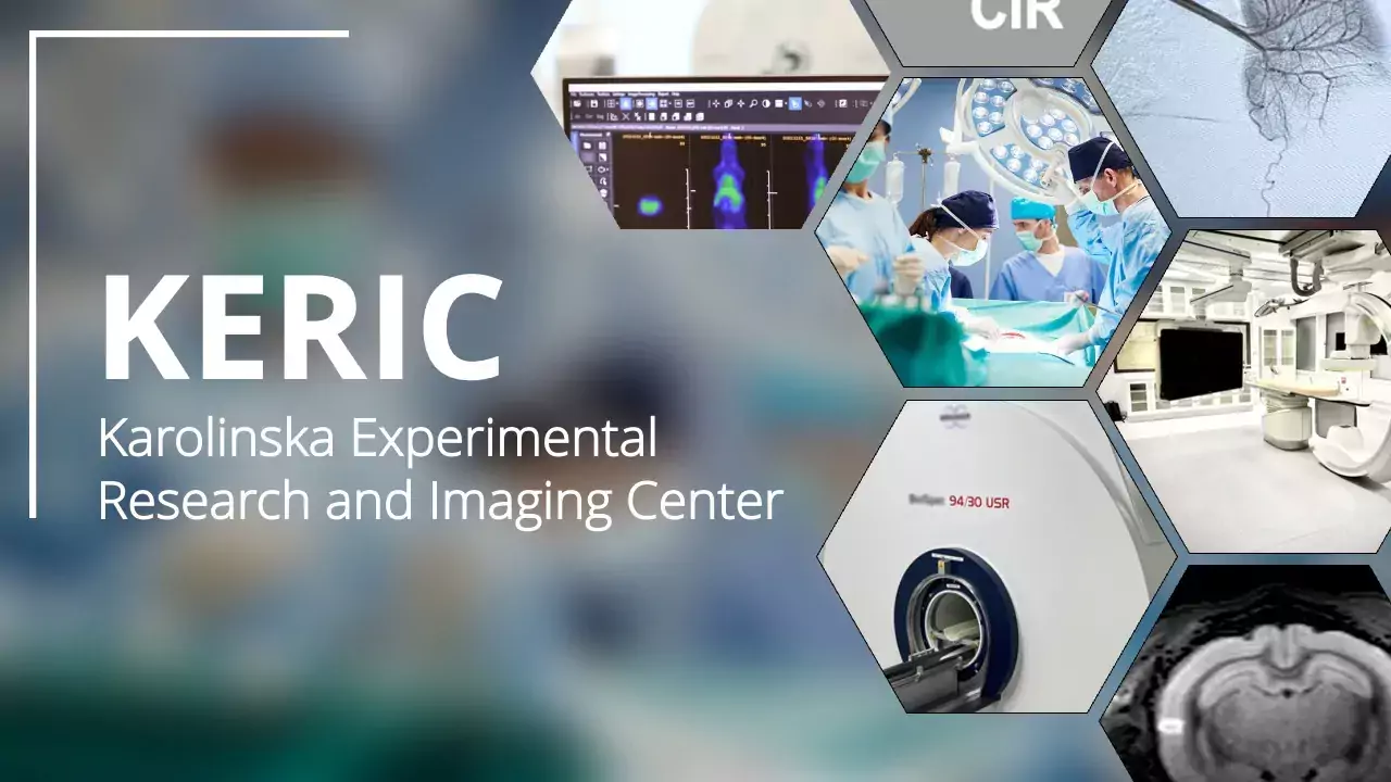 Photo: n/a
Menu for this area
Units at KERIC
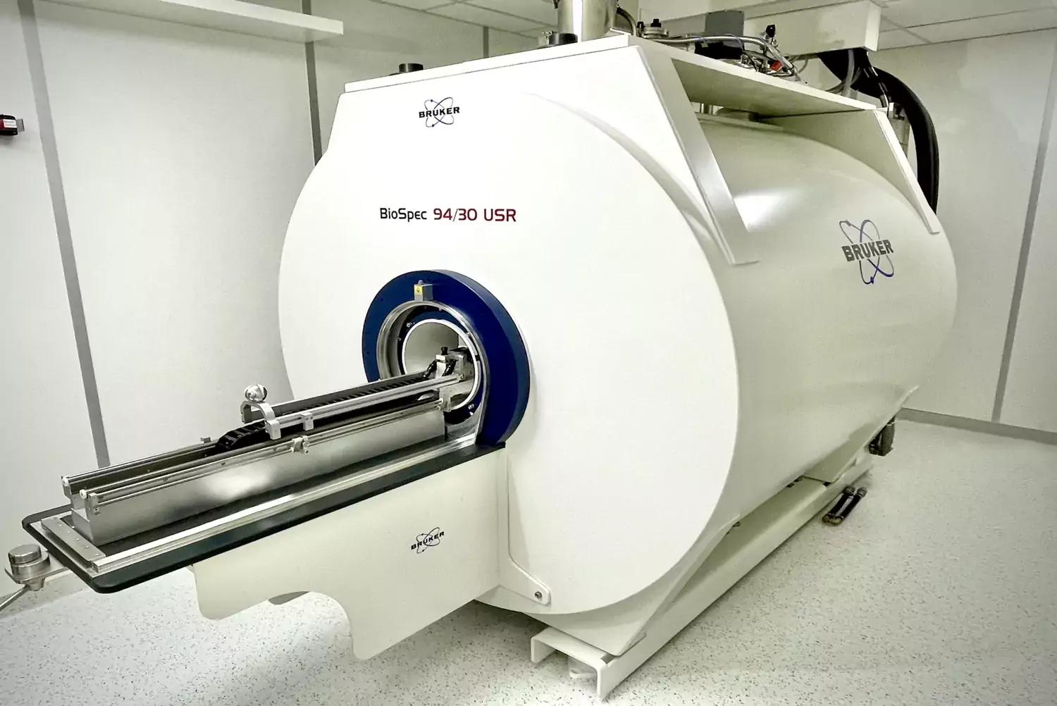 Photo: n/a
Preclinical MRI 9.4T
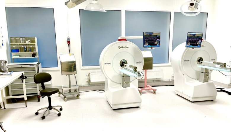 Photo: patjar
PET-CT/MR
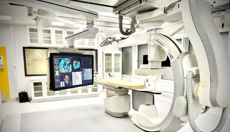 Photo: n/n
Angio CT
 Photo: patjar
Experimental Surgery
 Photo: n/a
Selected Research at KERIC
Some of our most successful customers that uses KERIC for their research.
 Photo: n/a
Example of Workflow
How we can help in different stages of a research project. Not just images.
 Photo: n/A
News for KERIC
https://ki.se/keric webpage update 2023-07-13
Selected projects and publications (scroll right)
Publications list from projects that have used KERIC facilities or collaborated direct with KERIC. List is in chronological order. Starting with the latest at top.
Please include KERIC in the acknowledgement, in publications based on results obtained from the any of our modalities or collaburated direct with us.
“The ***** was performed at the Department of Comparative Medicine/Karolinska Experimental Research and Imaging Centre at Karolinska University Hospital, Solna, Sweden.”
/Thank you

COVID-19 pathophysiology may be driven by an imbalance in the renin-angiotensin-aldosterone system
SARS-CoV-2 uses ACE2, an inhibitor of the Renin-Angiotensin-Aldosterone System (RAAS), for cellular entry. Studies indicate that RAAS imbalance worsens the prognosis in COVID-19. We present a consecutive retrospective COVID-19 cohort with findings of frequent pul- monary thromboembolism (17%), high pulmonary artery pressure (60%) and lung MRI perfusion disturbances. We demonstrate, in swine, that infusing angiotensin II or blocking ACE2 induces increased pulmonary artery pressure, reduces blood oxygenation, increases coagulation, disturbs lung perfusion, induces diffuse alveolar damage, and acute tubular necrosis compared to control animals. ...
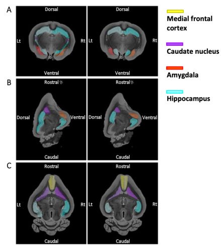
Changes in brain architecture are consistent with altered fear processing in domestic rabbits
The most characteristic feature of domestic animals is their change in behavior associated with selection for tameness. Here we show, using high-resolution brain magnetic resonance imaging in wild and domestic rabbits, that domestication reduced amygdala volume and enlarged medial prefrontal cortex volume, supporting that areas driving fear have lost volume while areas modulating negative affect have gained volume during domestication. ...
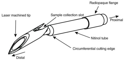
Myocardial micro-biopsy procedure for molecular characterization with increased precision and reduced trauma
Endomyocardial biopsy is a valuable tool in cardiac diagnostics but is limited by low diagnostic yield and significant complication risks. Meanwhile, recent developments in transcriptomic and proteomic technologies promise a wealth of biological data from minimal tissue samples. To take advantage of the minimal tissue amount needed for molecular analyses, we have developed a sub-millimeter endovascular biopsy device, considerably smaller than current clinical equipment, and devised a low-input RNA-sequencing protocol for analyzing small tissue samples. ...
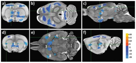
Functional connectivity in reward-related networks isassociated with individual differences in gambling strategies inmale Lister hooded rats
The aim of this explorative study was to examine individual strategies in the rGT and investigate possible behavioural and neural correlates associated with gambling strategies. ...
... R-fMRI results revealed associations between gambling strategies and brain regions central for reward networks. Moreover, rats with risky gambling strategies differed from those with strategic and intermediate strategies in brain functional connectivity. No differences in behavioural profiles, as assessed with the MCSF and novel cage tests, were observed between the gambling strategy groups. ...
