Technological platforms, Center for Infectious Medicine (CIM)
The Center for Infectious Medicine (CIM) has invested a significant amount of economic resources to amass cutting edge technology and scientific expertise. CIM offers advanced equipment for visualization and quantification of immune cell functions, as well as advanced flow cytometry. Researchers at CIM also have developed methods for isolating large numbers of specific primary cells and experimental In vivo models.
Imaging at the cellular and tissue level
To perform complete studies of immunological and pathogenic mechanisms, it is essential to have advanced equipment for visualization and quantification of immune cell functions.
CIM holds advanced equipment for imaging analyses including:
Light microscope equipped with specific acquired computerized image analyses programs for quantification of biological and immunological mediators.
Nikon A1R confocal microscope, which is an excellent tool for image analyses at the cellular and tissue level, including studies of immune cell functions, viral and bacterial entry into host cells, as well as intracellular vesicular transport and diffusion mechanisms. The microscope also provides imaging of live human immune cells and host pathogen interactions.
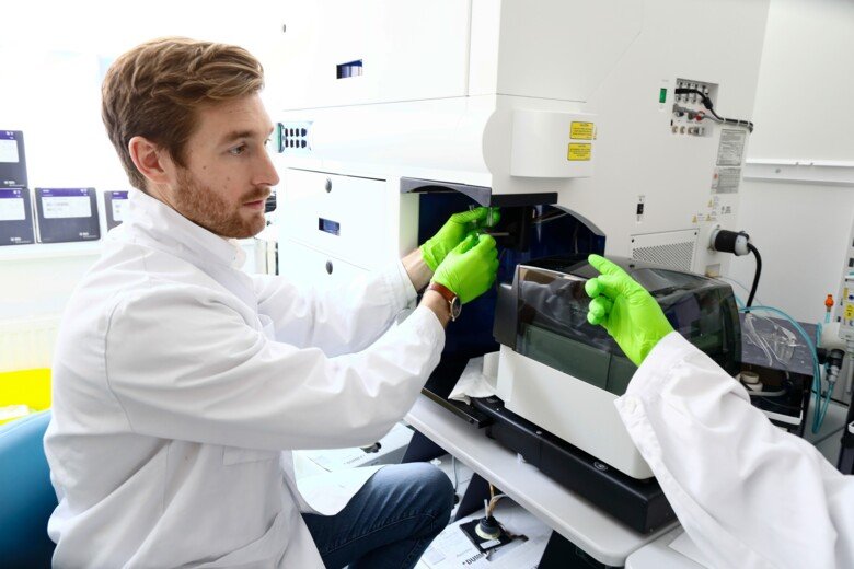
MACSima™ (High content imaging analyses), which allows detailed analyses on the expression levels of hundreds of protein markers within the spatial framework of tissues. The MACSima System is a fully automated instrument that utilizes fluorescence microscopy.
Tissue model systems
At CIM, researchers develop specialized cultures of tissue cells as well as ex vivo explant models. Several approaches are used to engineer three-dimensional (3D) tissue models, so-called organotypic cultures that also contain immune cells. Moving from cell monolayers to three-dimensional (3D) cultures is motivated by the need to work with cellular models that mimic the functions of living tissues.
For more information contact:
Mattias Svensson
Principal ResearcherAdvanced flow cytometry
This technology allows characterization of cells at single cell level and at high resolution, maximizing in-depth analysis of patient samples.
CIM is currently equipped with five flow cytometry analysis instruments:
- BD Symphony A5 (5 lasers, 29 fluorescence detectors)
- BD Symphony A3 (5 lasers, 28 fluorescence detectors)
- BD LSR Fortessa (5 lasers, 18 fluorescence detectors)
- BD Accuri (2 lasers, 4 fluorescence detectors)
- Cytek Aurora (5 laser spectral flow cytometer with capacity to detect >45 fluorophores)
In addition to the analysis instruments, there are two Sony MA900 cell sorters (2 lasers, 12 fluorescence detectors).
The Sony MA900 cell sorters are available for users at ANA Futura, enabling sorting of live cells in both BSL2 and BSL3 environment for downstream applications. The instruments are currently used by many groups in the environment in research related to human immune cell function and infectious diseases.
For more information contact:
Jakob Michaëlsson
Principal Researcher
Isolation of human immune cells from tissues
To study functional aspects of human immune cells it is crucial to have efficient methods of obtaining primary human cells. Researchers at CIM have developed several sophisticated methods for isolating specific subsets of primary cells. As an example, our researchers now isolate rare differentiated immune cell subsets and hematopoietic progenitor cells (HPC) from various tissues including human cord blood, gut, lung and liver.
For more information contact:
In vivo models
Experimental models constitute an important complement to in vitro experiments and studies on patient material.
Researchers at CIM use experimental models with an aim to gain increased understanding for complex host-microbe interactions, disease mechanisms and for testing new vaccine- and drug candidates. CIM is also actively involved in the generation of needs-driven, novel in vivo models for studies within these and related research areas.
10X Genomics single cell partitioning for DNA, RNA and protein analyses
To study molecular aspects of human immune cells at the single-cell resolution, CIM offers advanced instruments such as 10X Genomics single cell partitioning for DNA, RNA and protein analyses.