
Translational Cardiovascular Imaging – David Marlevi's team

The translational cardiovascular imaging team combines advanced medical imaging with bioengineering-driven image analysis to address urgent clinical challenges in cardiovascular care, as well as use novel imaging techniques to advance our understanding of cardiovascular disease.

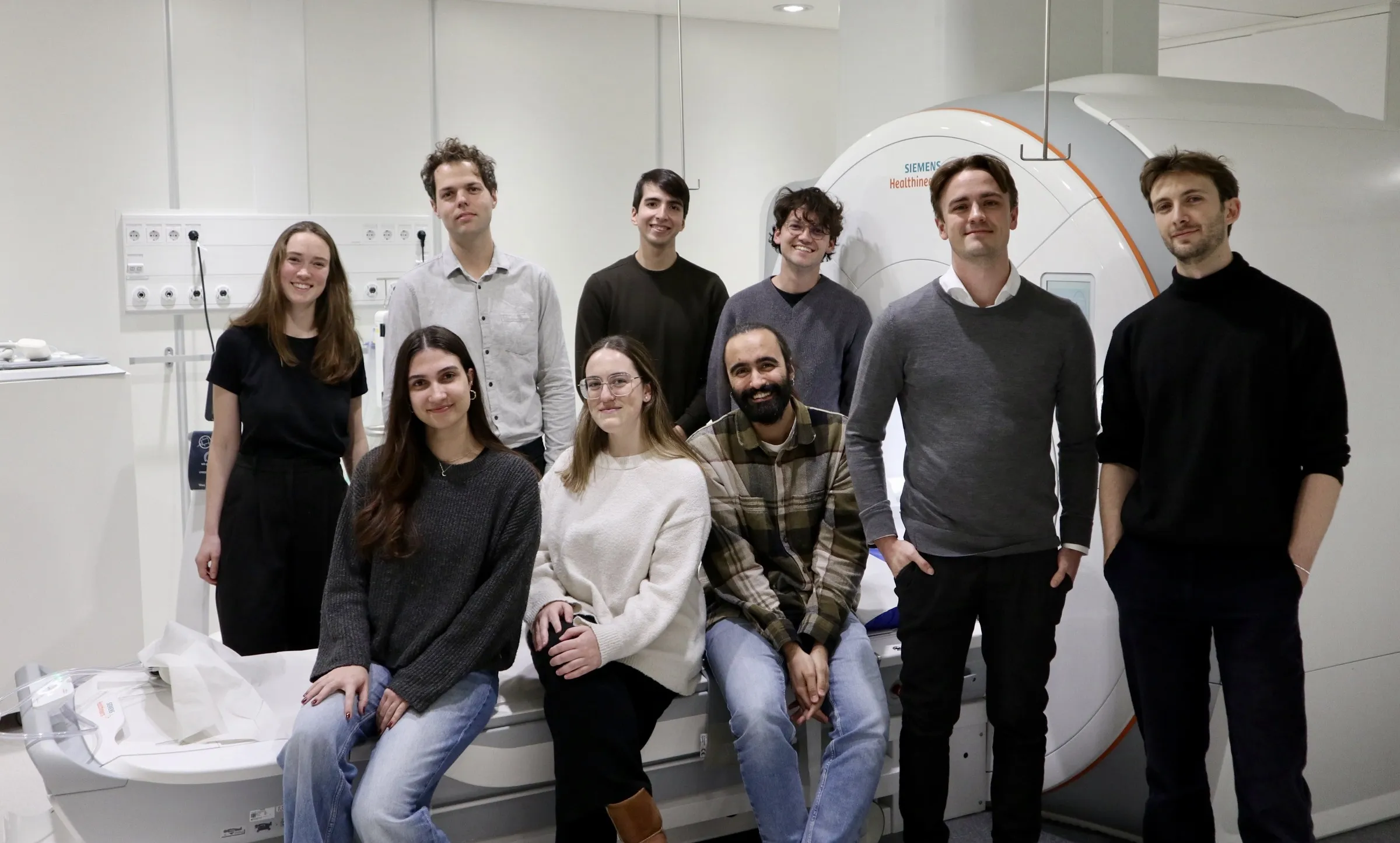
Nine members in front of a MRI scanner

Calendar

Our research

Cardiovascular disease remains the leading cause of death worldwide. Medical imaging plays a central role in cardiovascular disease management, enabling clinicians ad scientists to visualize and quantify both structural and functional abnormalities of the heart and vasculature. Further, the integration of advanced biomedical engineering tools (machine learning; computational modelling; bioinformatics) with medical imaging is starting to fundamentally change what we can image, and what we can quantify with clinical imaging systems.  

Led by David Marlevi, our research seeks to develop and implement image-based utilities to better diagnose, predict, and mechanistically understand cardiovascular disease. Being tightly connected to numerous collaborators across basic to clinical sciences, as well as with close connection to the Karolinska University Hospital, the team continuously seeks to produce impacting translational work with direct clinical relevance. 

Research vignettes 

Non-invasive estimation of cardiovascular pressure gradients

Regional quantification of cardiovascular pressure gradients is critical for diagnosis, treatment planning, and risk prediction of many cardiovascular disease. Still, for a large number of conditions, non-invasive assessment is obstructed by inherent method limitations, and a wide range of clinical instances exist where regional pressure behaviour remains unexplored. To tackle this, and funded by the European Research Council, we have deployed a combination of physics-informed image analysis (invoking fundamental fluid mechanical description of blood flow) and full-field flow imaging (4D Flow MRI) to allow for arbitrary probing of pressure gradients across previously inaccessible compartments. Through this work, we seek to extend these utilities to further understand early hemodynamic changes indicative of later physiological impairement, including validation, implementation, and clincial utility across spatial (large / small vessels), temporal (fast / slow flows) and flow (laminar / turbulent) scales.

Super-resolution MRI 

The advent of full-field flow MRI has fundamentally changed our ability to interrogate complex hemodynamic behaviour in a direct clinical setting. However, spatiotemporal limitations exist based on the clinical time frames in which the systems can be used, obstructing assessment of regional or highly transient flow events. To tackle this, we have employed a variety of trained neural networks to enhance spatial image resolution and image quality, effectively pushing quantitative volumetric MRI into challenging vascular beds and flow domains.

Imaging the biology of atherosclerosis 

Stroke remains a leading causes of mortality worldwide, with identification of unstable carotid plaques key for improved diagnosis. While lesion instability has been coupled to generic phenotypes, the spatial interplay between plaque morphology, biomechanics, and biology remains unknown. To address this, and in direct collaboration with the Vascular Surgery Group, we are now integrating state-of-the-art imaging, plaque biobank data, lesion-specific modelling, and spatial transcriptomics to uncover the regional interplay between spatial plaque morphology, biomechanics, and biology, working towards individualized plaque risk predictions. 

Non-invasive risk prediction of chronic aortic dissection

Aortic dissection (AD) involves the sudden delamination of the aortic wall and formation of a false lumen (FL). While most AD patients survive the acute phase, approximately 50% of AD patients who survive to hospital discharge will either die of aorta-related causes or require surgical repair within a decade – complications thought to arise primarily due to the progressive growth of the FL. Predicting which AD patients are at high risk of FL growth is thus critically important. In this project, we are developing and utilizing a combination of 4D MRI and CTA to non-invasively map FL bio- and hemodynamics, seeking to shed new light on the complex pathophysiology of progressive dissection growth.  

Publications

All publications from group members

Funding

  • European Research Council
  • Swedish Research Council
  • Swedish Heart Lung Foundation
  • Ulla Hamberg Angeby and Lennart Angeby's Foundation
  • Knut and Alice Wallenberg Foundation / Data-Driven Life Science
  • MedTechLabs (together with PI: Ulf Hedin (KI) and Christian Gasser (KTH))
  • Stockholm City Council (ALF)
  • Magnus Bergvalls stiftelse

Staff and contact

Group leader

All members of the group

Team alumni

  • Sebastian Jofré
  • Leon Ericsson
  • Adam Hjalmarsson
  • Simon Åslund
  • David Bardvall
  • Meritxell Pardo Rel 
  • Raquel Riera Turiel

Contact and visit us

Postal address

K1 Molekylär medicin och kirurgi 
K1 MMK Klinisk fysiologi
SE-171 76 Stockholm

Visiting address

Eugeniavägen 3, Karolinska University Hospital, A8:01, 17176 Stockholm

Map to Eugeniavägen 3