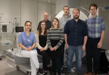
Quantitative Cardiovascular Physiology – Jannike Nickander's team

We combine quantitative cardiac magnetic resonance (CMR) imaging with proteomic and biomarker analysis to understand coronary microvascular dysfunction and ischemic heart disease in patients without obstructive coronary artery disease. By integrating advanced imaging biomarkers with omics data, we aim to improve diagnostic precision and uncover the mechanisms behind these common but underdiagnosed conditions, translating findings into better-targeted clinical care.

Our research

Coronary microvascular dysfunction (CMD) and myocardial infarction with non-obstructive coronary arteries (MINOCA) are increasingly recognized causes of chest pain and ischemic heart disease, yet remain underdiagnosed — particularly in women. Quantitative cardiac MRI provides a unique, non-invasive window into myocardial perfusion, tissue characterization, and microvascular function, allowing us to detect disease mechanisms that conventional angiography cannot capture.

Led by Jannike Nickander, our research combines state-of-the-art quantitative CMR (including stress perfusion imaging, T1/T2 mapping, and extracellular volume quantification) with proteomic profiling to build a more complete picture of the pathophysiology underlying CMD and MINOCA. We are a part of the research group for Clinical Physiology at Karolinska Institutet and Karolinska University Hospital, and collaborate across cardiology, radiology, and laboratory medicine to bridge mechanistic imaging findings with clinical translation.

Research vignettes

Coronary microvascular dysfunction and quantitative stress perfusion CMR

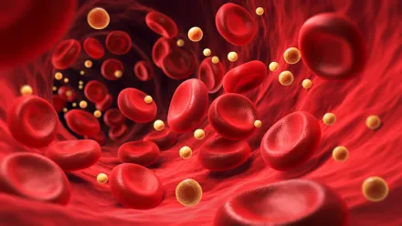
Coronary microvascular dysfunction is a key contributor to ischemic symptoms in patients with angina but no obstructive coronary artery disease. Using quantitative stress perfusion CMR, we characterize microvascular blood flow reserve non-invasively, aiming to refine diagnostic criteria and better identify patients at risk of adverse cardiovascular outcomes.

Sex differences in cardiac imaging biomarkers

Women are disproportionately affected by MINOCA and CMD, yet diagnostic imaging thresholds have historically been derived from male-predominant cohorts. Our work has demonstrated sex differences in myocardial blood volume, perfusion, and extracellular volume at rest and during vasodilator stress, providing mechanistic insight that is now informing sex-specific reference values in quantitative CMR.

Follow-up and long-term outcomes in MINOCA

Patients with myocardial infarction with non-obstructive coronary arteries often lack a clear mechanistic diagnosis at the time of the acute event. Through comprehensive follow-up CMR, we investigate the evolution of myocardial injury, microvascular function, and clinical outcomes in this population to guide more precise long-term management.

Integrating proteomics with cardiac imaging

By combining high-throughput proteomic platforms (e.g., Olink) with quantitative imaging biomarkers, we aim to identify circulating protein signatures associated with microvascular dysfunction and myocardial tissue changes, opening avenues for novel non-invasive biomarkers and a deeper mechanistic understanding of ischemic heart disease without obstructive coronary disease. 

Publications

All publications from group members

Funding

  • Swedish Heart Lung Foundation
  • Swedish Society of Medicine
  • Region of Stockholm (ALF)
  • Karolinska Institutet
  • Magnus Bergvalls stiftelse

Staff and contact

Group leader

All members of the group

Alumni

  • Raman Vashishtha
  • Abdumalek Al-Hashimi
  • Rebecka Vesterlund
  • Ivan Bekhali
  • Esra Mohamed
  • Bilal Blbisi 

Contact 

For more information, research opportunities, student projects, or collaborations please contact the team leader:

Jannike Nickander; jannike.nickander@ki.se

Postal address

K1 Molekylär medicin och kirurgi 
K1 MMK Klinisk fysiologi
SE-171 76 Stockholm

Visiting address

Eugeniavägen 3, Karolinska University Hospital, A8:01, 17176 Stockholm