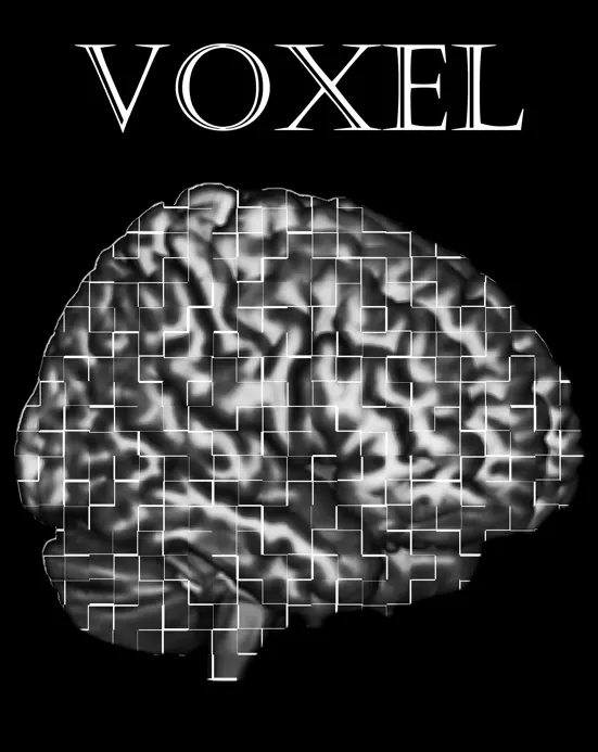
The VOXEL group at ARC
We are specialised in neuroimaging techniques and methods pertaining to magnetic resonance imaging and positron emission tomography.

Focus
We have a strong focus on novel imaging techniques and methods, and multimodal imaging.
Aim
We want to contribute to the development of neuroimaging techniques and methods and create opportunities to establish new collaborations.
What we do
Here are some examples of what we currently do.
- Brain iron imaging using quantitative susceptibility mapping and signal separation (paramagnetic/diamagnetic)
- MR Elastography to assess stiffness and viscoelastic properties of the brain tissue and multidimentional diffusion, in collaboration with KTH Royal Institute of Technology
- PET imaging of the dopaminergic system, in collaboration with Umeå University, University or Turku, Finland
- Development of a high-resolution T2 sequence on 3T MRI for the assessment of the olfactory bulbs in collaboration with SUBIC, Stockholm University
- Development of a deep learning tool for the automated segmentation of midbrain nuclei (see section Available codes)
- Contributions to the Stanford Multilayer Integration of Networks Toolbox (see section Available codes)
- MR spectroscopy in collaboration with SUBIC and Örebro University
Current group members
Farshad Falahati
Research Infrastructure Specialist, EngineerGrégoria Kalpouzos
Associate Professor, Senior LecturerMartin Nilsson
Doctoral studentJavier Oltra
Postdoctoral ResearcherGoran Papenberg
Associate Professor, Principal ResearcherBárbara Avelar
Assistant ProfessorAlireza Salami
Associate Professor, Principal ResearcherFrida Smids
Doctoral student| MRI | Summary measures/assessment |
|---|---|
| T1-weighted | Morphometrics, perivascular spaces |
| Diffusion Tensor Imaging (DTI) | White-matter integrity, structural connectome |
| Functional MRI (task, rest) | Activation, functional connectome |
| T2-FLAIR | White-matter hyperintensities |
| Multi-echo Gradient Recalled Echo (meGRE) | Relaxometry R2*, Quantitative Susceptibility Mapping (QSM) for iron quantification |
| Fast-Spin Echo, Magnetization Transfer | Neuromelanin (locus coeruleus, substantia nigra) |
| High-resolution T2 | Olfactory bulbs' anatomy |
| MR Elastography | Cerebral stiffness, viscosity-elasticity |
| Multidimensional diffusion (MUDI) | Subvoxel diffusion metrics (e.g., microanisotropy) |
| Pseudo-continuous Arterial Spin Labeling (pCASL) | Quantitative cerebral blood flow (perfusion) |
| Susceptibility Weighted Imaging (SWI) | Microbleeds |
| Neurite Orientation Dispersion and Density Imaging (NODDI) | Neurite Density Index, Orientation Dispersion Index |
| 1H MR Spectroscopy (MEGA-)PRESS, semi-LASER | Metabolite concentrations (e.g., NAA, Cr, Cho, mI, Glx, GABA) |
| PET | |
|---|---|
| 11C-Raclopride | Dopamine D2 receptors (at rest, and under reward) |
| 11C-SCH23390 | Dopamine D1 receptors |
| 18F-DOPA | Dopamine synthesis |
| 18F-PE2I | Dopamine transporter availability |
| 11C-PBR28 | TSPO density, microglia |
We also make use of publicly available data such as florbetapir (AV-45) and flortaucipir (AV-1451) PET for amyloid and tau, respectively.
Reference: Falahati F. Gustavsson J, Kalpouzos G (2024). Automated segmentation of midbrain nuclei using deep learning and multisequence MRI: A longitudinal study on iron accumulation with age. Imaging Neuroscience, 2: 1-20. Doi: 10.1162/imag_a_00304
Reference: Sarraf S, Avelar-Pereira B, Hosseini SMH; Alzheimer’s Disease Neuroimaging Initiative (2025). Multilayer Integration of Networks Toolbox (MINT). Communications Biology 8(1):894. doi: 10.1038/s42003-025-08269-4.
Contact
Grégoria Kalpouzos
Senior LecturerContact us if you have any questions or would be interested in joining the group.
