3D-EM Core Facility
The 3D electron microscopy core facility (3D-EM) is a new facility for high-end biomedical electron microscopy at Solna campus. The facility is equipped with (cryo) TEMs, cryo FIB-SEM, cryo confocal, plunge freezing, and high-pressure freezing instruments for single particle analysis and tomography. Contact us to discuss a project and start training on our equipment.
Menu for this area
Main areas
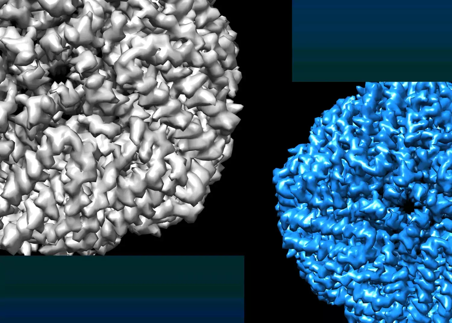 Photo: Amy Bondy
Single particle cryo EM
Single particle cryo-EM enables 3D reconstruction of individual proteins and biological complexes through collection of micrographs of purified sample, followed by 2D class averaging, and 3D reconstruction.
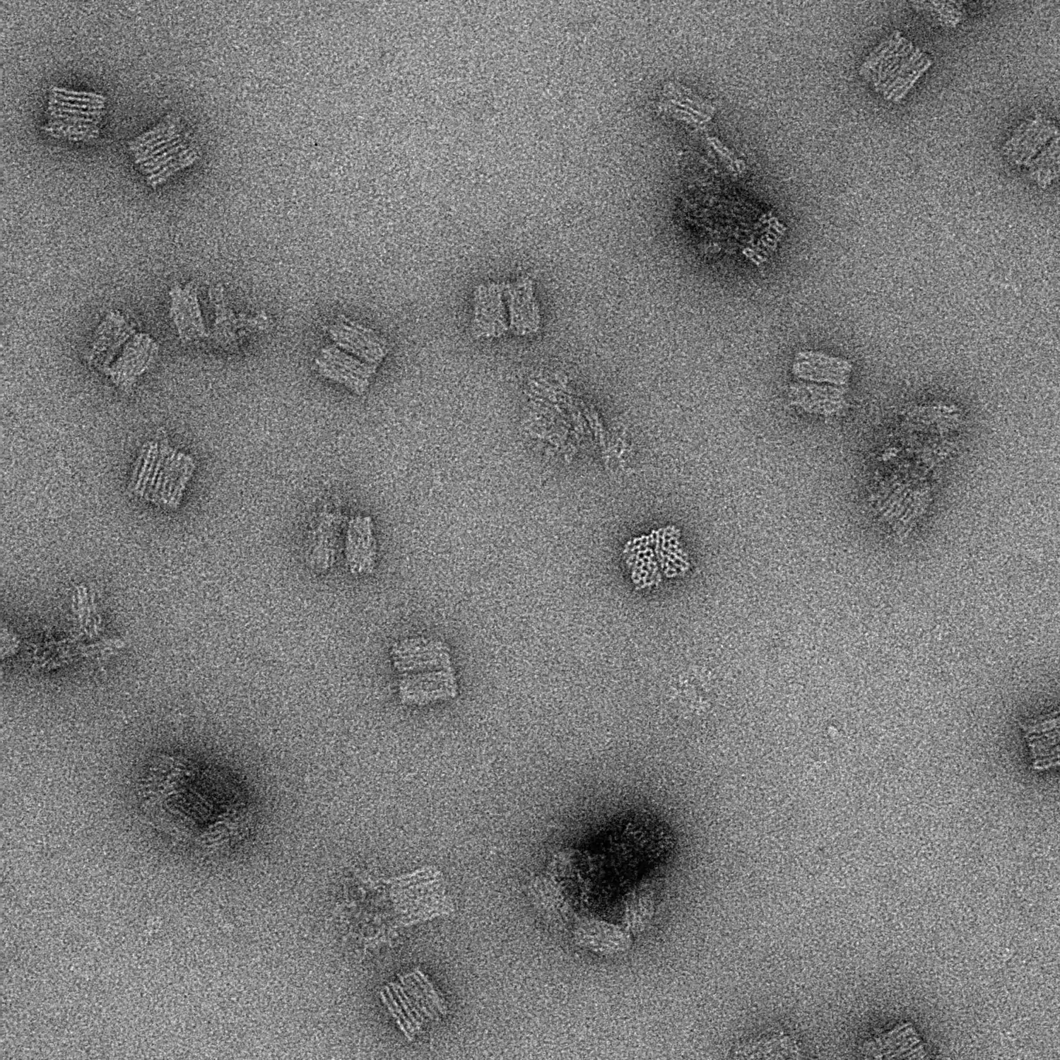 Photo: Amy Bondy
Negative stain EM
Negative staining is a procedure which embeds small biological proteins such as proteins, protein complexes, or nanoparticles on a TEM grid in a thin film of heavy metal salts (i.e. uranyl salts) to enhance contrast and reveal their structural details. Unlike cryo-TEM, this process is carried out at room temperature.
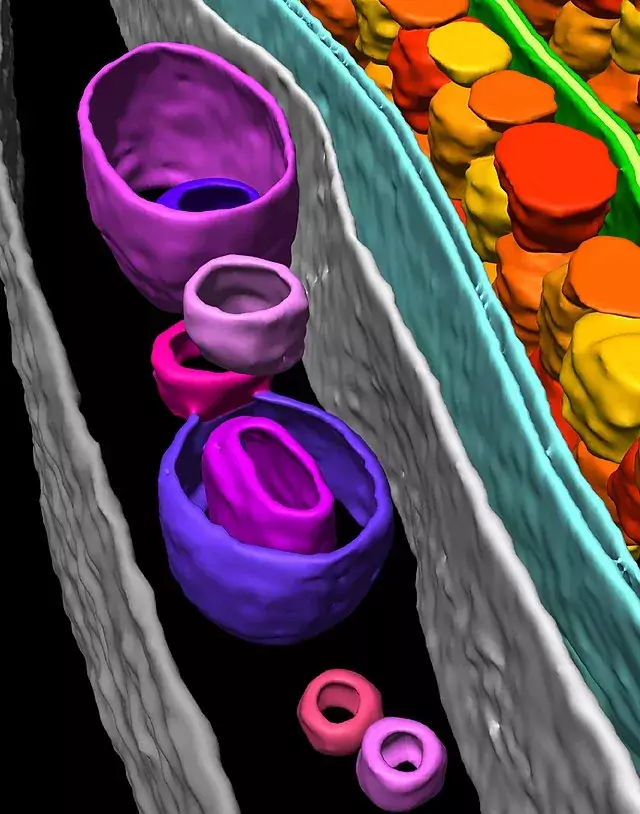 Photo: Wikepedia Commons
Tomography
Electron tomography enables visualization of proteins and organelles in situ; within the cell without isolation and purification. This method can be used under cryo conditions for cells, or at room temperature to look at fibers or other non-symmetrical particles.
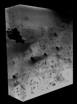 Photo: Amy Bondy
Volume imaging
Volume imaging electron microscopy, also called slice-and-view, reveals the 3D ultrastructure of cells and tissues through continuous depths of at least 1 micrometer. Volume imaging generates a series of images of the specimen, that, when combined, form a digital representation of the specimen volume.
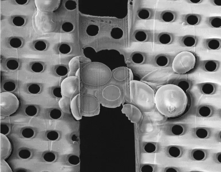 Photo: Amy Bondy
Lamella milling
Because cells and tissue samples are often too thick to image directly with cryo electron tomography, lamella milling is an important preparation step that thins the sample until it is electron-beam transparent (approximately 200 nm thick).
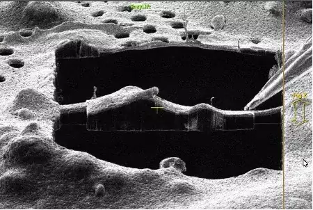 Photo: Amy Bondy
FIB milling with liftout
This preparation technique enables cryo-electron FIB milling and tomography to be performed on cells from multicellular organisms or tissue samples, rather than just single cells, extending the range of applications for in situ structural biology.
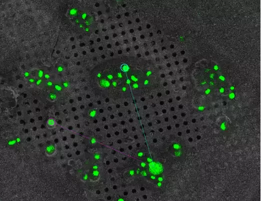 Photo: Amy Bondy
Cryo-CLEM
Cryo correlative light and electron microscopy (CLEM) consists of locating areas of interest using cryo-fluorescence light microscopy, followed by inspecting the sample at high resolution using electron microscopy. This provides protein localization of individually labelled biomolecules, which can then be targeted for lamella milling and/or tomography.
 Photo: Pixabay_gef24fc607
Related publications
Publications resulting from 3D-EM Core Facility use.
 Photo: Genís Valentín Gesé
Facts about our core facility
Location: Wargentin, Nobels väg 12, Campus Solna
Director: Martin Hällberg
Key words: electron microscopy, structural biology, TEM, SEM, single particle, tomography
Department: Department of Cell and Molecular Biology
Funding: Karolinska Institutet, user fees
External use: 3D-EM is also available to external users (find out more about External use of KI's core facilities)
 Photo: Lasse Skog