Volume Imaging
Volume imaging electron microscopy, also called slice-and-view or serial block-face imaging, reveals the 3D ultrastructure of cells and tissues through continuous depths of at least 1 micrometer. Volume imaging generates a series of images of the specimen, that, when combined, form a digital representation of the specimen volume.
Explore
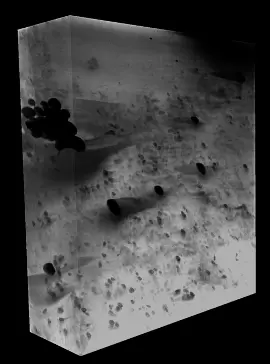
Volume imaging using a dual-beam focused ion beam/scanning electron microscope (FIB/SEM; Aquilos 2) enables large volumes of material to be reconstructed into 3D maps, for imaging cells and tissues. The individual images are acquired by repeatedly slicing the specimen into thin sections using the FIB, and then imaging the exposed face of the sample with the SEM. Volume imaging enables imaging and assessment of subcellular structures on the mesoscale (10 nm to 10 µm), compared to tomography which images at a much higher magnification. Although lower resolution, the lower magnifications used with volume imaging have the advantage of achieving an overall picture of a cell or tissue sample. Following image acquisition, data processing steps include alignment, filtering to remove image artifacts, and segmentation.
The 3D-EM facility specializes in cryo volume imaging, but plastic-embedded room-temperature projects can also be performed on our equipment.
The workflow can be conducted using the 3D-EM facility’s: EM ICE ► sp8 cryo confocal ► Aquilos 2
Data collection softwares available: Slice-and-View
Processing softwares available: Amira
Additional resources
Volume EM: a quiet revolution takes shape.
Collinson LM, Bosch C, Bullen A, Burden JJ, Carzaniga R, Cheng C, Darrow MC, Fletcher G, Johnson E, Narayan K, Peddie CJ, Winn M, Wood C, Patwardhan A, Kleywegt GJ, Verkade P
Nat Methods 2023 Jun;20(6):777-782
Cryo-plasma FIB/SEM volume imaging of biological specimens.
Dumoux M, Glen T, Smith JLR, Ho EML, Perdigão LMA, Pennington A, Klumpe S, Yee NBY, Farmer DA, Lai PYA, Bowles W, Kelley R, Plitzko JM, Wu L, Basham M, Clare DK, Siebert CA, Darrow MC, Naismith JH, Grange M
Elife 2023 Feb;12():