Single particle cryo EM
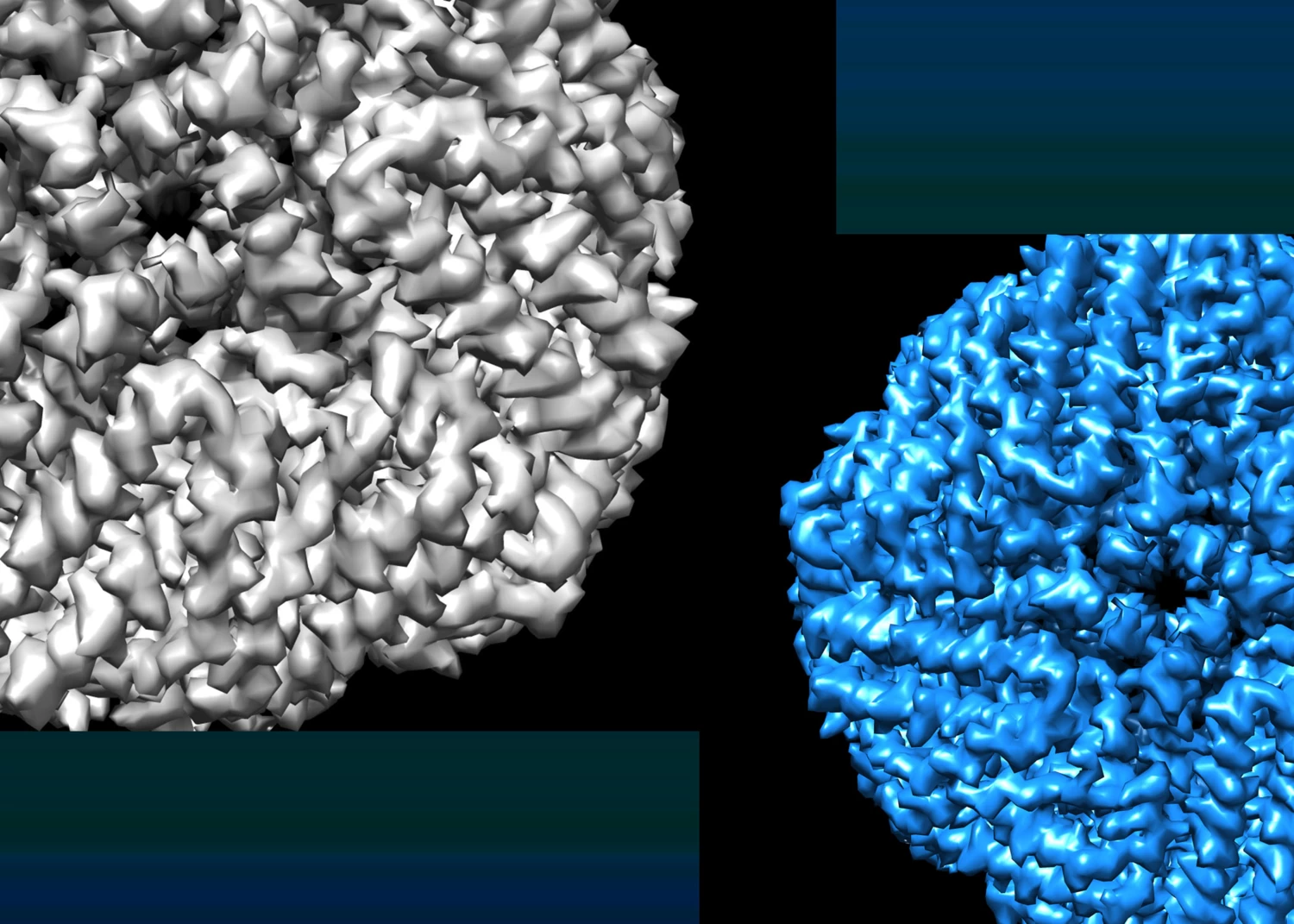
Single particle cryo-EM enables 3D reconstruction of individual proteins and biological complexes through collection of micrographs of purified sample, followed by 2D class averaging, and 3D reconstruction.
Explore
Single particle analysis of cryo biological samples is a standard cryo-EM technique that allows you to investigate the structure of biomolecules at near-atomic resolutions, achieving resolutions up to 2 Å.
In single particle analysis, purified proteins or protein complexes are suspended in vitreous ice through plunge freezing (Vitrobot), which preserves the protein’s native structure. Our liquid nitrogen cooled TEM (Krios) is then used to semi-automatically collect thousands of micrographs, each containing multiple particles oriented randomly within the ice.
Data processing is then conducted on the output micrographs to average particles with the same orientation and reconstruct the protein’s high resolution 3D structure.
The workflow can be conducted using the 3D-EM facility’s: Vitrobot ► Krios TEM
Data collection softwares available: EPU, Serial EM
Processing softwares available: WARP
Additional resources
Single-particle cryo-electron microscopy.
Doerr A
Nat Methods 2016 Jan;13(1):23
What Could Go Wrong? A Practical Guide to Single-Particle Cryo-EM: From Biochemistry to Atomic Models.
Cianfrocco MA, Kellogg EH
J Chem Inf Model 2020 May;60(5):2458-2469
A basic introduction to single particles cryo-electron microscopy.
Raimondi V.; Grinzato, A
AIMS Biophysics 2022, Volume 9, Issue 1: 5-20