TOUCH TO TRIUMPH Stain smarter, Discover faster
We invite the scientific community to a series of presentations and workshops under the theme 'Stain smarter, discover faster'.
Presentations and workshops
Date: 16 Oct
Time: 11am-12pm
Place: Biomedicum: Eva & Georg Klein
Fully automated spatial omics staining platform enabling: High throughput and large staining area (30 slides per run)
Standardization and reproducibility of staining protocols
High sensitivity utilizing signal amplification and multiplexing
High precision and fast speed (2–3 hours per run)
Fully open system with customizable protocols
Capability to perform assays like RNAScope, SignalStar, Akoya Opal, and other multiplex IHC/IF/FISH assays
Multiplex IHC and IF with Tyramide Signal Amplification (TSA)
Cyclic multiplex IF with iterative rounds of staining
Pre-analytical workflows for spatial transcriptomics, including:
- Region of interest (ROI) mapping
- Antibody validation for platforms including 10x Genomics Xenium, MACSima, GeoMx DSP, and PhenoCycler
Date: 21 Oct
Time: Drop-in
Place: Biomedicum: B0517
Registered participants can submit tissue slides and antibodies and receive results in 3 hours
Date: 21 Oct
Place: Biomedicum: Nils Ringertz
- Multiplexing and spatial biology by Cell Signaling Technologies and Vector Laboratories
Time: 11.45 am
Pick up your lunch
- “Accelerate Spatial Biology: Rapid, High-Precision Multiplex IHC with SignalStar™ by Cell Signaling Technologies
Time: 12 pm-1 pm
Abstract: Accelerate discoveries in spatial biology by 70% with SignalStar™ Multiplex IHC, which offers optimized panels for efficient results in just two days and eliminates time-consuming optimization and validation processes. Join our seminar led by our Senior Global Product Trainer and discover this innovative technology that enhances spatial biology research by amplifying cellular protein signals in FFPE tissue samples. Gain rapid insights, visual clarity with up to 8 (or more!) target detections, enhanced precision, immediate start with validated protocols, and seamless integration into existing fluorescence imagers with SignalStar Multiplex IHC.
Speaker info: Dr. Theisgen completed a Diploma in Biochemistry and a PhD in Molecular Medicine at the University of Halle (Germany). She joined CST in 2018 as Manager for the European Technical Support Team and also worked as a Field Application Scientist for two years, where she gained extensive technical knowledge of the CST product portfolio.
In 2022, she took over the position as Global Product Trainer. Since then, she has conducted numerous technical trainings for customers, but also for distribution partners worldwide. Her experience with various antibody applications enables her to support customers in their research with troubleshooting, target selection, protocol development and data interpretation.
- “Next-Generation Reagents for Reliable Multiplex IHC/IF” by Vector Laboratories
Time: 1pm-2pm
Abstract: Vector Laboratories, with a 50-year legacy, is a global leader in immunohistochemistry, immunofluorescence, glycobiology, and bioconjugation. Our innovative products, including the VECTASTAIN® Elite ABC Kit, VectaShield® Antifade Mounting Medium, and TrueVIEW® Autofluorescence Quenching Reagent, are widely used in research, cited in over 350,000 publications. We now introduce VectaPlex™ and Glysite™ Explorer. VectaPlex™ enables efficient antibody removal from FFPE tissue sections without damaging tissue or antigens. Glysite™ Explorer offers a comprehensive kit for spatial glycan detection near proteins, supporting advanced studies of protein glycosylation in various sample types.
Speaker info: Deborah Purvis brings over 28 years of experience in the medical diagnostics industry, with a strong foundation in diagnostic research gained through her work at the Wellcome Foundation and several leading Universities across the UK. Throughout her career, Deborah has held diverse roles across multiple companies in the UK, Europe, and Globally. In recent years, she has focused on advanced technologies at Vector Laboratories, specializing in immunohistochemistry (IHC) and immunofluorescence (IF), and is currently the Director of Channel Management and Business Development for the company in Europe, Middle East, Africa and India.
In addition to her professional work, Deborah is proud to serve as Chair of the Board of LabXCell, the organization that oversees UK NEQAS Cellular Pathology Technique. This independent body is responsible for providing external quality assurance for cellular pathology techniques across the UK, Europe, and Internationally.
Date: 23 Oct
Time: 11 am-2:30 pm
Place: Biomedicum: Eva & Georg Klein
With Niklas Peterson, Visiopharm
Niklas Petersen is a trusted expert in digital pathology and image analysis, helping researchers and clinicians unlock the full potential of AI-powered workflows. At Visiopharm, he specializes in translating complex spatial biology data into clear, actionable insights—whether through intuitive demos, tailored consultations, or hands-on collaborations.
With a passion for making high-tech approachable, Niklas is the go-to person for turning tissue images into meaningful data—one cell at a time.
Abstract: Quantifying RNA in tissue sections is critical for understanding disease mechanisms, particularly in cancer research and diagnostics. However, translating RNA signals into meaningful biological insights can be challenging. Staining inconsistencies, overlapping cells, and signal ambiguity often complicate analysis — especially when high precision is required.
In this seminar, we will demonstrate how Visiopharm’s AI-powered image analysis platform enables robust and reproducible analysis of RNAscope data. By incorporating both nuclear and membrane stains, our platform goes beyond traditional nucleus-based segmentation to accurately identify individual cells, even in complex environments such as tumor microenvironments or densely packed tissues.
Join us to see how advanced AI models can help overcome the limitations of manual interpretation and transform scattered RNA "spots" into high-quality spatial data — unlocking deeper insights into cell-level expression, localization, and disease biology.
Date: 23 Oct
Time: 1 pm - 2.20 pm
Place: Biomedicum: Eva & Georg Klein
Live Demo at the Workshop
Bring your challenges and image analysis questions! We’ll host a live demo to show how you can maximize your output using Visiopharm’s flexible platform.
You’ll also get hands-on exposure to Visiopharm’s interactive data exploration and plotting tools. These include t-SNE, scatter and box plots, co-occurrence matrices, and cell galleries—all tightly integrated into the workflow. These tools let you explore cellular phenotypes visually, adjust thresholds in real time, and link every plotted data point back to its corresponding cell in the image for quick QC and verification
Bonus: One participant will be selected as a winner during the workshop — and receive a personalized deep-dive session on their data.
Deadline for abstract submission: October 13
Submit Your Abstract — and Get 2–3 Images analysed by Niklas!
Working with RNAscope, IHC, or multiplexed tissue data? Submit your abstract for a chance to win a personalized image analysis session with Niklas Petersen.
Using Visiopharm’s AI-powered tools, Niklas will demonstrate how to transform your data into meaningful, quantitative insights — tailored to your specific challenges.
Spots become science — let’s make your data shine!
by Melissa Turan ACDBio
Date: 28 Oct
Time: 11 am-12 pm seminar
1 pm-2 pm lunch
Place: Biomedicum: Nils Ringertz
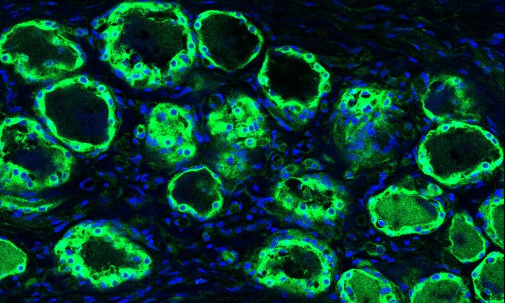
RNAscope is a revolutionary in situ hybridization (ISH) assay that detects target RNA within intact cells, delivering both high sensitivity and high specificity via a proprietary “double Z” probe design that amplifies true signals while suppressing background noise It enables single-molecule RNA detection, visualized as individual punctate dots under a microscope, bringing together spatial context and molecular precision. The technology is trusted widely, with applications across various species, tissues, and research areas, and has been cited in over 10,000 publications—a testament to its robustness and reproducibility
Date: 28 oct
Time: 1:30-2:30 pm
Place: Biomedicum Room: B0317
Abstract
Quantifying and contextualizing biomarker expression within tissue is essential for understanding cellular behaviour while maintaining the integrity of complex tissue architecture. In this session, Dr. Ghislaine Lioux will demonstrate how Indica Labs’ HALO image analysis powerhouse unites a wide range of quantitative analyses in a single, intuitive platform requiring no technical expertise. With HALO and HALO AI, tissue can be compartmentalized and cells segmented consistently for robust, reproducible analysis. This presentation will highlight how HALO enables precise cell-level quantification, scoring and phenotyping of RNAscope and Multiplex IF data. Combined with HALO Link, it creates a complete 360° digital pathology ecosystem where results can be easily reviewed, seamlessly shared, and trends effortlessly identified.
Hands-on session
HALO Hands-On Workshop: Experience AI-Powered Image Analysis for RNAscope and Multiplex IF
Join Indica Labs for an interactive HALO Hands-On Session led by Dr. Ghislaine Lioux, Senior Field Application Scientist. In this guided workshop, participants will explore the power and simplicity of HALO for quantitative tissue image analysis. Using demo datasets on provided laptops, attendees will learn how to:
- Use AI-driven annotation and Tissue classification,
- Perform cell-based quantification of protein and/or RNA signal
- Visualize data trends and share results through HALO Link
Whether you’re new to HALO or looking to enhance your digital pathology workflows, this session offers a practical, hands-on introduction to extracting meaningful insights from your own data.
Speaker Bio
Dr. Ghislaine Lioux is a Senior Field Application Scientist at Indica Labs. In her role, Ghislaine assists scientists in academia, healthcare, and pharma worldwide to turn complex multiplex and RNA in situ data into high-impact discoveries through HALO, HALO AI, and HALO Link. With over a decade of experience in immunostaining and imaging across Europe and the USA, Ghislaine combines deep scientific expertise with a passion for enabling researchers to extract meaningful biological insights from tissue data. She earned her PhD from the Spanish National Institute of Cardiovascular Research in Madrid as a Marie Curie Fellow, where she studied molecular mechanisms of heart development.
Date: 29–31 Oct
Time: Drop-in
Place: Biomedicum Room: B0517
Registered participants can submit tissue slides and receive results overnight.
Special discounts: 75% off RNAScope LS kits and 50% off LS probes - ONLY BOND RX compatible". The discount is valid only during the event. For more information RNAscope on BOND RX
All participants will have access to imaging support at BIC (Biomedicum Imaging Facility).
Date: 6 nov
Time: 11 am
Place: Biomedicum Room: B0333
VoxlBio – a KI Innovations DRIVE incubator company – will present their novel in situ genetic visualization technology that enables direct detection of gene expression and single-nucleotide mutations within intact tissue at single-cell resolution.
Abstract
VoxlBio introduces a groundbreaking targeted sequence-capture approach that preserves spatial context while delivering unparalleled sensitivity and specificity—distinguishing RNA molecules differing by as little as a single nucleotide. This platform opens new opportunities in advanced diagnostics, targeted therapy development, and fundamental disease research.
We warmly invite all registered participants to join this additional session and discover how VoxlBio’s innovation can accelerate precision medicine and spatial biology research.