Negative stain EM
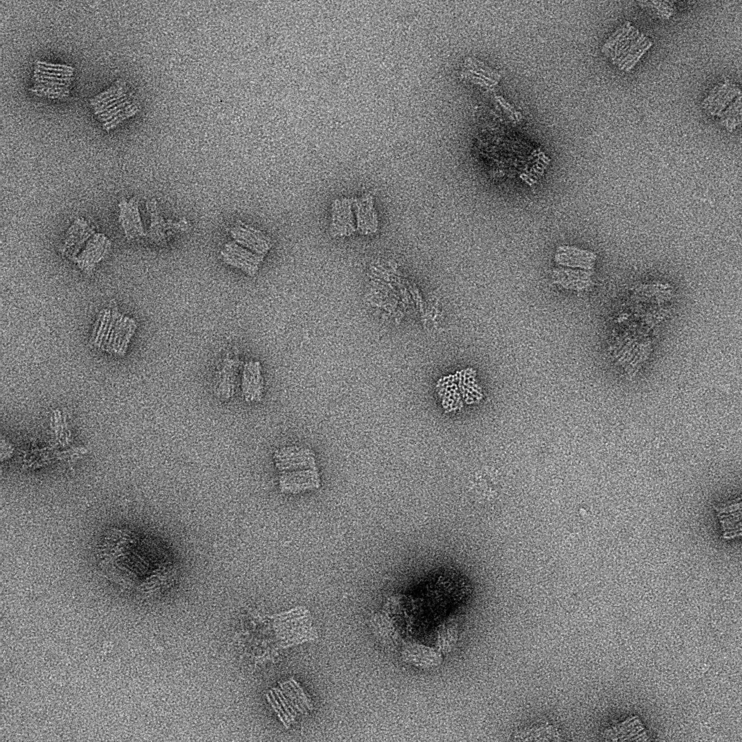
Negative staining is a procedure which embeds small biological molecules such as proteins, protein complexes, or nanoparticles on a TEM grid in a thin film of heavy metal salts (i.e. uranyl salts) to enhance contrast and reveal their structural details. Unlike cryo-TEM, this process is carried out at room temperature.
Explore
Negative staining for biological particles is a standard EM technique that allows you to investigate the size and structure of biomolecules and nanoparticles, achieving resolutions up to 20 Å.
Negative staining is often used in conjunction with cryo-EM to check the sample before freezing, to ensure that the protein complex is intact, the sample is not aggregated, and the concentration is appropriate. Negative staining can also be used independently from cryo-EM to determine a 3D structure.
Traditionally, uranyl acetate or uranyl formate are used for staining followed by room temperature TEM (Talos). Unless you have electron-dense nanoparticles (such as gold), a heavy metal stain is necessary to visualize the relatively electron-translucent materials.
In this process, particles are surrounded by the stain, which scatters electrons strongly creating negative contrast, whereby the particle appears bright against the stain. Parameters such as staining time and blotting technique affect the proportions between negative and positive staining on the grid and may need to be optimized for each sample.
The workflow can be conducted using the 3D-EM core facility’s: Talos
Data collection softwares available: Tia or Velox (for manual image collection/screening), Serial EM
Additional resources
Visualizing proteins and macromolecular complexes by negative stain EM: from grid preparation to image acquisition.
Booth DS, Avila-Sakar A, Cheng Y
J Vis Exp 2011 Dec;(58):
Variations on Negative Stain Electron Microscopy Methods: Tools for Tackling Challenging Systems.
Scarff CA, Fuller MJG, Thompson RF, Iadanza MG
J Vis Exp 2018 Feb;(132):
Negative and Positive Staining in Transmission Electron Microscopy for Virus Diagnosis.
Barereto-Vieira, D.F.; Barth, O.M.
Microbology in Agriculture and Human Health (2015).