Lamella milling
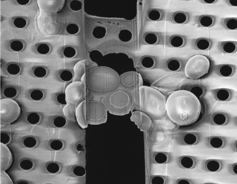
Because cells and tissue samples are often too thick to image directly with cryo electron tomography, lamella milling is an important preparation step that thins the sample until it is electron-beam transparent (approximately 200 nm thick).
Explore
Used either alone (Aquilos 2) or following correlative microscopy (Leica sp8 cryo confocal), the focused ion beam is used to prepare a thin, electron-transparent lamella by removing material above and below the target region. This lamella contains the region of interest and can be milled as thin as 100–200 nanometers.
There is no mechanical sectioning; instead, the vitrified sample is thinned with the help of a focused beam of gallium ions that is scanned across the frozen sample surface, removing surface atoms in a layer-by-layer fashion (called sputtering or ion beam milling).
Sample thinning is essential for the tomography workflow because the electron beam in the TEM can only pass through samples that are thin enough to transmit 200–300 keV electrons. Cryo-FIB thinning is a straightforward and more manageable method compared to cryo-ultramicrotomy, and it avoids intrinsic cutting artifacts of mechanical sectioning at cryo-temperatures.
Lamella can be milled either manually or the process can be automated in the software AutoTEM.
The workflow can be conducted using the 3D-EM facility’s: Vitrobot ► Aquilos 2
Data collection software available: AutoTEM
Additional resources
Cryo-electron tomography on focused ion beam lamellae transforms structural cell biology.
Berger C, Premaraj N, Ravelli RBG, Knoops K, López-Iglesias C, Peters PJ
Nat Methods 2023 Apr;20(4):499-511
Waffle Method: A general and flexible approach for improving throughput in FIB-milling.
Kelley K, Raczkowski AM, Klykov O, Jaroenlak P, Bobe D, Kopylov M, Eng ET, Bhabha G, Potter CS, Carragher B, Noble AJ
Nat Commun 2022 Apr;13(1):1857