FIB milling with liftout
This preparation technique enables cryo-electron FIB milling and tomography to be performed on cells from multicellular organisms or tissue samples, rather than just single cells, extending the range of applications for in situ structural biology.
Explore
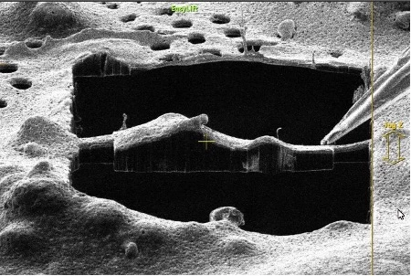
FIB lift-out is used for site-specific preparation of high-quality TEM samples from bulk materials, such as from tissue samples or large cells from multicellular organisms.
In this method, a focused ion beam (Aquilos 2) is used to remove material around a site of interest, forming a lamella. The lamella is then extracted from the bulk sample, transferred to a TEM grid with a cryo-cooled nanomanipulator (EasyLift), and thinned to electron transparency for tomography. The original, thick sample is typically high pressure frozen (Leica EM ICE) rather than plunge frozen. Correlative microscopy (Leica sp8 cryo confocal) can also be used to target the location of the target of interest before milling.
This workflow can be conducted using the 3D-EM facility’s: EM ICE ► sp8 cryo confocal ► Aquilos 2
Data collection software available: AutoTEM
Additional resources
A cryo-FIB lift-out technique enables molecular-resolution cryo-ET within native Caenorhabditis elegans tissue.
Schaffer M, Pfeffer S, Mahamid J, Kleindiek S, Laugks T, Albert S, Engel BD, Rummel A, Smith AJ, Baumeister W, Plitzko JM
Nat Methods 2019 Aug;16(8):757-762
Cryo-FIB milling and lift-out for preparation of specimens for cryo-TEM.
Zachman, M.J.; Noble J.M.; Kourkoutis, L.F.
Microscopy and Microanalysis 23, 2312-2313 (2017).
Serial lift-out—Sampling the molecular anatomy of whole organisms.
Schiotz, O.H.; et al.
BioRxiv (2023).