Cryo-CLEM
Cryo correlative light and electron microscopy (CLEM) consists of locating areas of interest using cryo-fluorescence light microscopy, followed by inspecting the sample at high resolution using electron microscopy. This provides protein localization of individually labelled biomolecules, which can then be targeted for lamella milling and/or tomography.
Explore
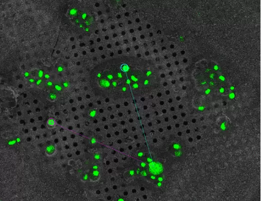
Cryo-correlative light and electron microscopy (cryo-CLEM) combines the advantages of both fluorescence light microscopy as well as 3D cryo-electron tomography to reveal the ultrastructure of significant target molecules with specific cellular functions.
Cryo-CLEM allows imaging of cells and tissues by means of fluorescence microscopy exhibiting the location of the molecule at high temporal and spatial resolution while tomography is used to analyze the 3D structure at a molecular resolution.
With a thin sample, tomography can be performed directly (Krios) following fluorescence imaging (sp8 cryo confocal). However if the sample is thick, the XYZ location can be determined and following correlation, either volume imaging or lamella milling can be performed (Aquilos 2) first.
This workflow can be conducted using the 3D-EM facility’s:
- Vitrobot ► sp8 cryo confocal ► Krios
- Vitrobot ► sp8 cryo confocal ► Aquilos 2 ► Krios
- EM ICE ► sp8 cryo confocal ► Aquilos 2 ► Krios
Data collection software available: Maps (used for correlation)
Additional resources
Advances in Cryo-Correlative Light and Electron Microscopy: Applications for Studying Molecular and Cellular Events.
Jun S, Ro HJ, Bharda A, Kim SI, Jeoung D, Jung HS
Protein J 2019 Dec;38(6):609-615
Recent advances and current trends in cryo-electron microscopy.
Guaita M, Watters SC, Loerch S
Curr Opin Struct Biol 2022 Dec;77():102484
Super-resolution confocal cryo-CLEM with cryo-FIB milling for in situ imaging of Deinococcus radiodurans.
Sexton DL, Burgold S, Schertel A, Tocheva EI
Curr Res Struct Biol 2022 ;4():1-9