FENO - our instruments
The unit has its own histological laboratory with advanced instruments for automatic multiplex immunohistochemistry, RNA in situ hybridization, and slide-scanner. Samples are registered individually using barcode to facilitate sample identification, data documentation, and digital scanning of sections. Imaging data is documented and digitalized using light, polarized and fluorescence slide scanner or multispectral fluorescence microscopy, followed by image analysis using different programs.
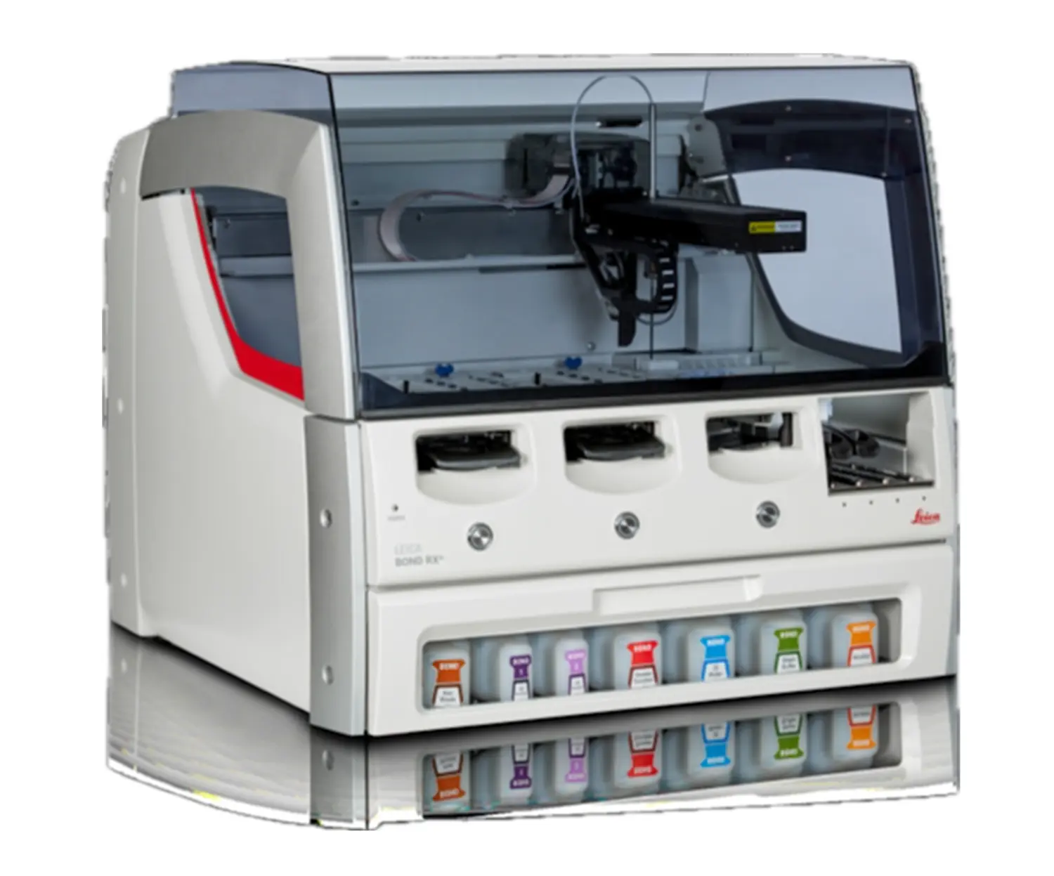
Leica BOND RXm fully automated research stainer
Deliver reproducible, quality slides and eliminate time spent on manual staining with the BOND RXm fully automated benchtop stainer for IHC, ISH, and emerging tests.
- Multiple choices of automat staining: IF, IHC, ISH, FISH, CTC, multiplexing
- Protocol customization upon request
- Reduce process variability
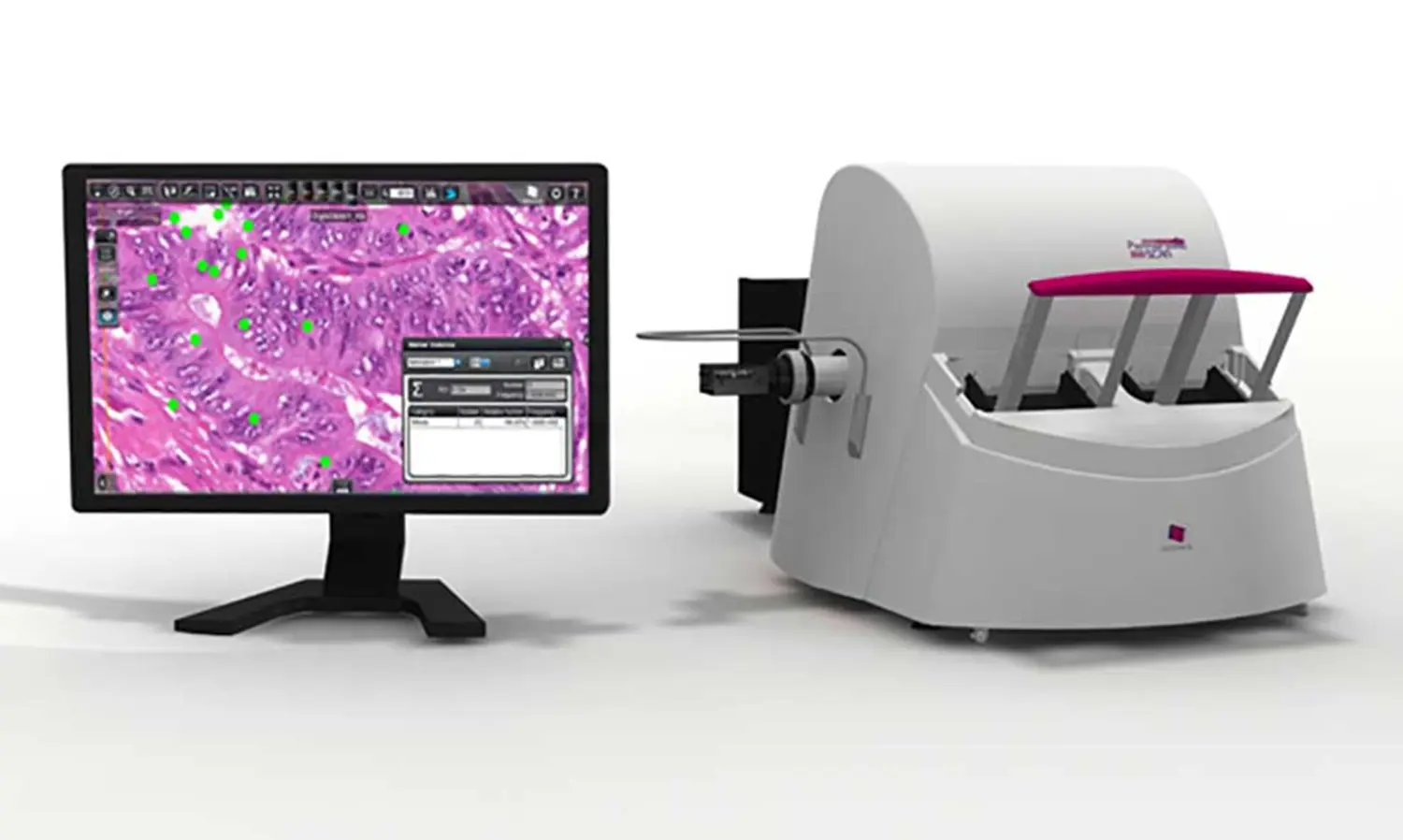
3D histech section scanner
The PANNORAMIC SCAN is a medium-capacity digital slide scanner by 3DHISTECH, with its 150-slide capacity and brightfield scanning abilities.
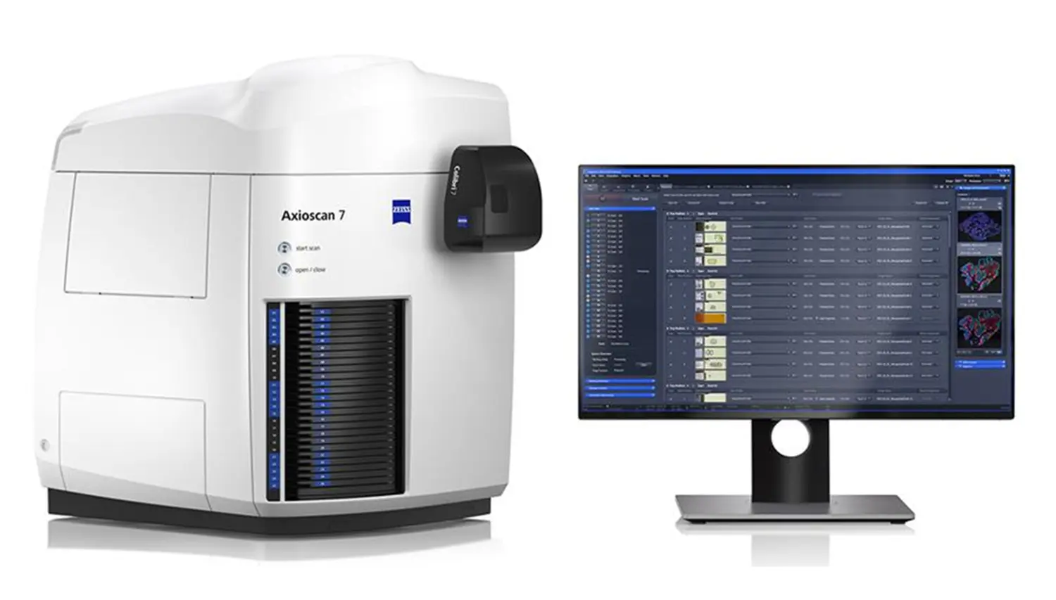
High-performance slide scanner ZEISS Axioscan 7
ZEISS Axioscan 7, an automatic microscopic scanner with bright field, polarization and fluorescence imaging function scanner, is a new available instrument with FENO in 2024. Axioscan 7 is equipped with high-performance optics from ZEISS, enabling high-resolution and detailed images of tissue samples. This is important to detect microscopic details and to facilitate diagnostic decisions. Axioscan 7 supports different sample sizes and can handle both thin sections and whole tissue sections. The device offers advanced image processing functions that allow to improve contrast, color reproduction and sharpness to optimize image quality. Additionally, digital images can be easily shared and archived for future reference.
- Fluorescence mode: 7 filters; 380 - 770 nm
- Brightfield mode
- Polarization mode
- Rapid switching between different modes
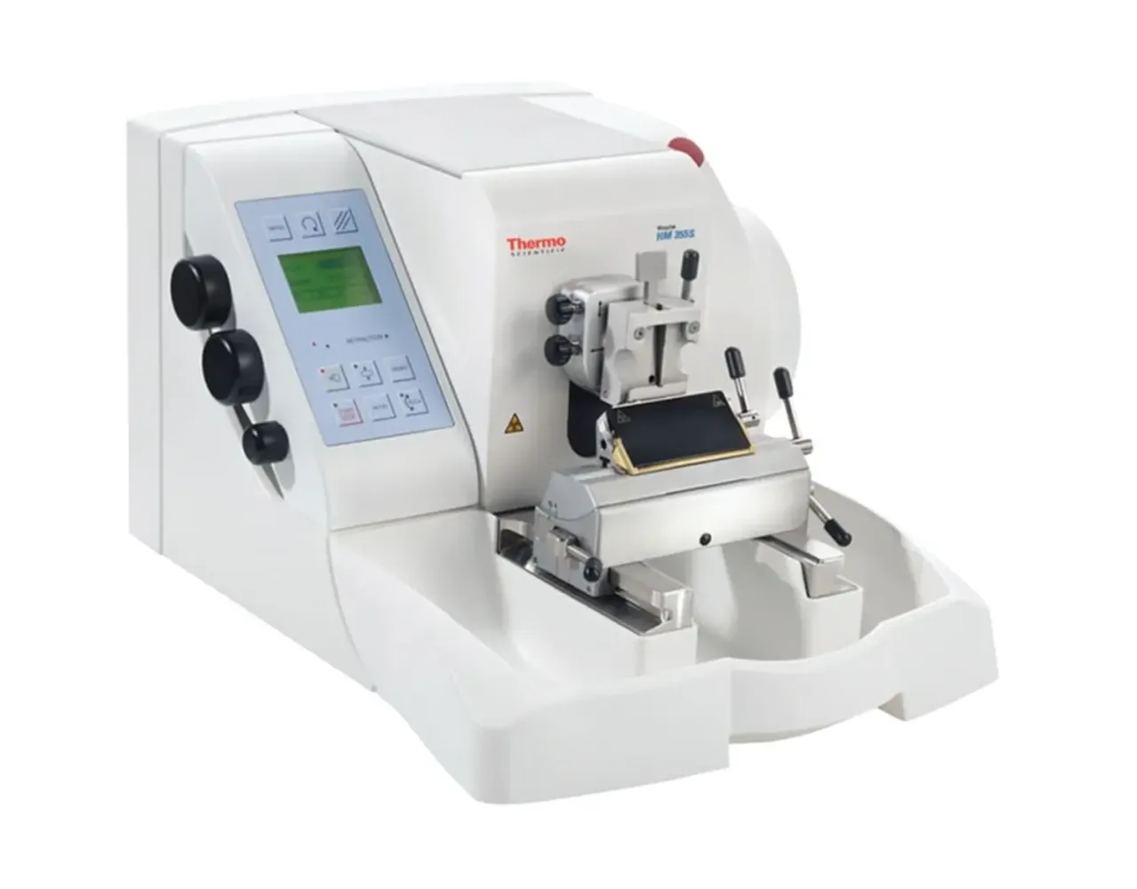
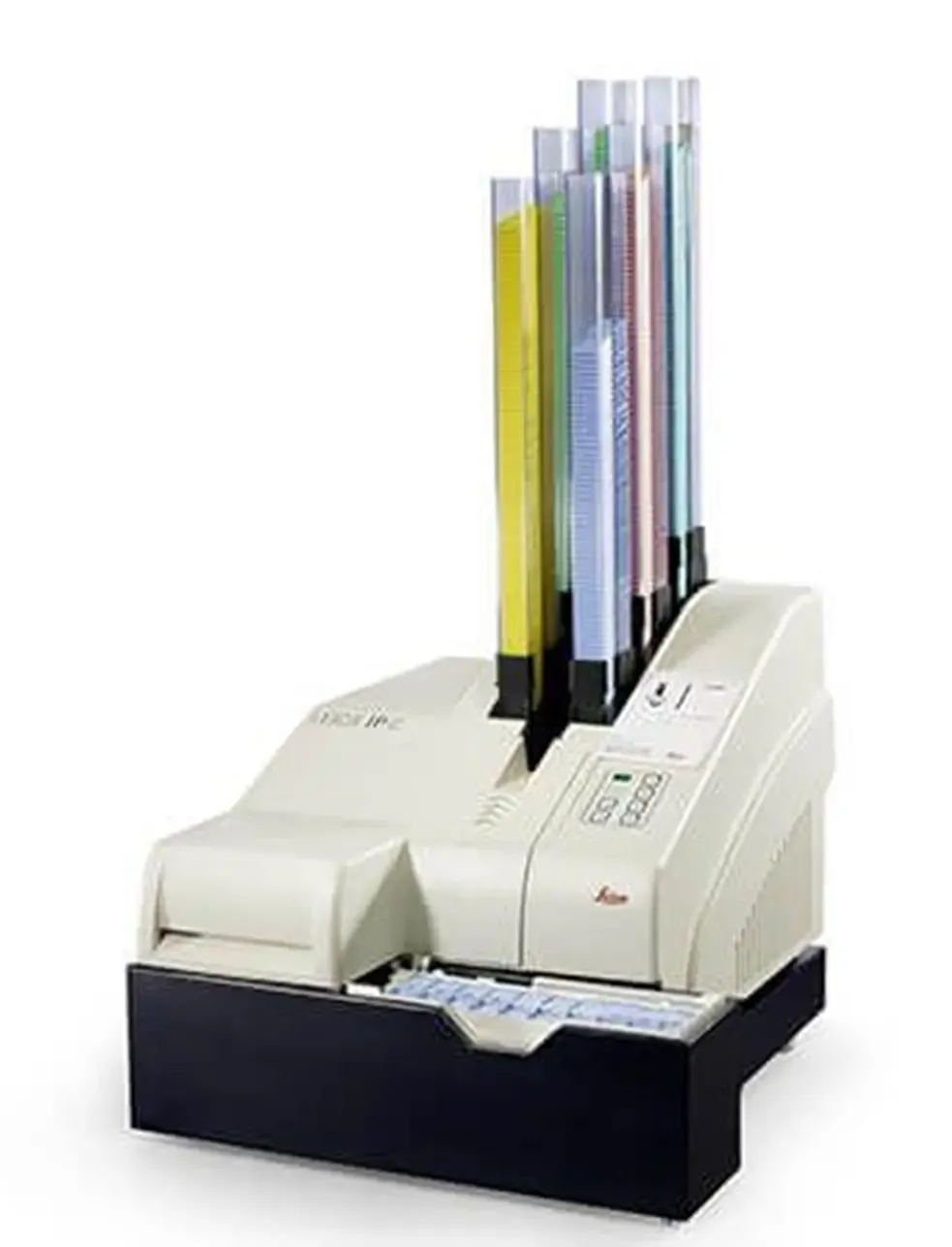
Additional instruments