Positron Emission Tomography (PET)
Positron Emission Tomography (PET) is an unsurpassed method for imaging biochemical and physiological processes in vivo.
PET imaging uses the rapid radioactive decay of positron-emitting isotopes (such as 11C, 15O, 18F and 68Ga), which are, after their generation in cyclotrons or elution generators, chemically incorporated in molecules with high biological relevance. These radiotracers are intravenously injected prior to the scan or after positioning in the PET scanner. Then, positrons are emitted and will annihilate with electrons within a very short time under the emission of two 511 keV photons. PET scanners utilize the coincidence detection of these photons with rings of detectors.
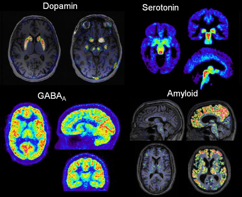
PET combined with tracer kinetic models measures ligand receptor interactions, membrane transport, metabolism and blood flow noninvasively and quantitatively in vivo. PET has been used extensively to study cellular metabolism and molecular interactions in the brain, heart and malignant tumors.