Science in Art, Art in Science. Lennart Nilsson and Karolinska Institutet
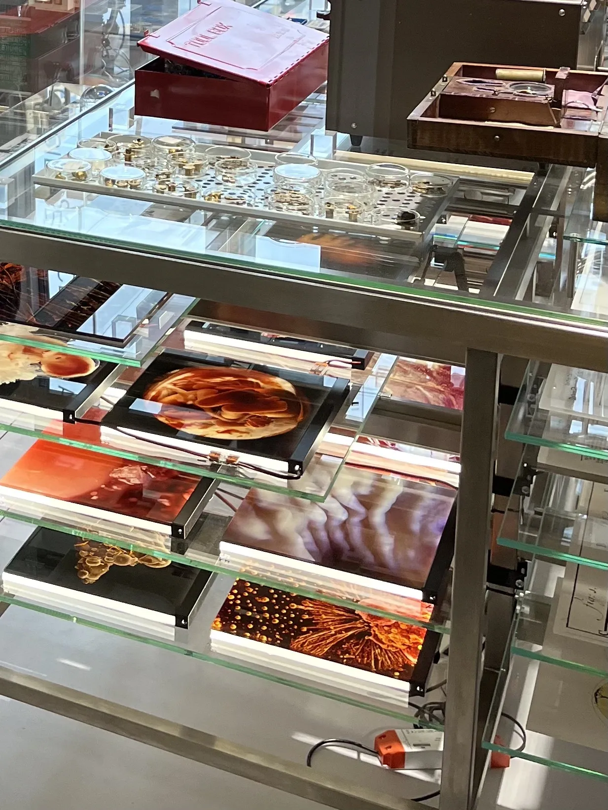
The exhibition Science in Art - Art in Science: Imaging Technologies and Lennart Nilsson at Karolinska Institutet shows unique images and objects from photographer Lennart Nilsson's many years of work at Karolinska Institutet. Nilsson brought an aesthetic vision to scientific images and was very result oriented. Time and again he pushed the technical boundaries of photography.
The exhibition is produced by the department for Medical History and Heritage at KI.
Visit the exhibition: Nobels väg 5, campus Solna.
To book a guided tour, contact Olof Ljungstrom.
The exhibition places Lennart Nilsson's work in a larger historical context. Instruments for seeing beyond the capabilities of the human eye were developed from the 16th century. Studies in optics improved telescopes and microscopes. In the 17th century, instruments for seeing the very distant, such as telescopes, and the very small; microscopes, were improved. As a precursor to Lennart Nilsson's images of reproduction, Antonie van Leeuwenhoek developed a new generation of microscopes at the end of the 17th century. Among his discoveries are sperm, the male sex cells.
Experiments and observations were used to investigate general principles about the world and nature. The microscope in particular provided knowledge about structure. In medicine, the discovery was made that all living things reproduce through cell division. The pathological side of this revealed how processes can go wrong and abnormal cell division lead to disease. During the decades around the middle of the 20th century, KI went through a phase where research was driven by the opportunity to find new answers by building its own technical solutions. At KI's new campus in Solna, one third of the premises were workshops. The researchers asked questions to a staff of engineers, technicians and craftsmen.
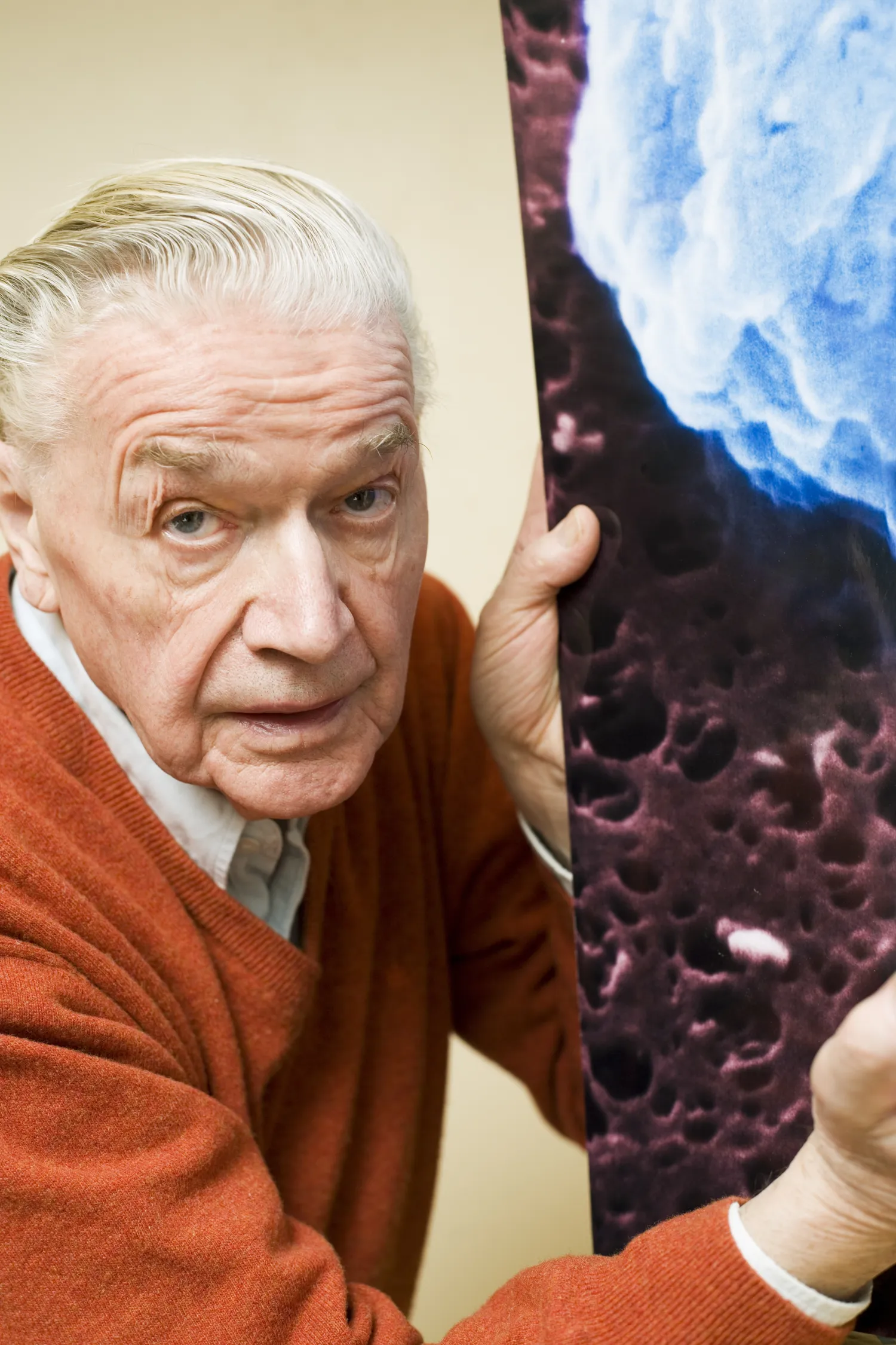
Lennart Nilsson moved around the clinics and laboratories of Stockholm in the 1950s, environments he came to depict. After an impressive career as a press photographer with a flair for pregnant images, he began a journey into the microcosm of the organic world. The culmination came with the publication of color photographs of fetuses seemingly floating in the womb, or space, published in Life Magazine, The Drama of Life Before Birth, April 30, 1965.
Lennart Nilsson took an even deeper step into the organic world with the electron microscope. If the 17th century discovered the sperm and the 19th century the egg, Lennart Nilsson was able to capture their fusion on image in 1990. Alongside reproduction and sexuality, Lennart Nilsson, like his predecessors, captured pathological phenomena. A sensation was the first electron microscopic image of the HIV virus. Lennart Nilsson also photographed SARS, bird flu, and many Corona viruses that infect both animals and humans.