Using AI to diagnose breast cancer earlier
Although mammography has dramatically reduced mortality rates for breast cancer, current technology still misses almost one third of cases. There is also a serious shortage of radiologists. Researchers believe that these problems can be solved by artificial intelligence (AI).
Text: Magnus Trogen Phalén, first published in Swedish in the magazine Medicinsk Vetenskap No 3/2020
The chances of surviving breast cancer are significantly higher if the disease is detected at an early stage. Studies demonstrate that those who have regular mammographs have a 40% lower risk of dying from breast cancer within 10 years of a diagnosis. Unfortunately, there is a serious shortage of radiologists to examine mammographs.
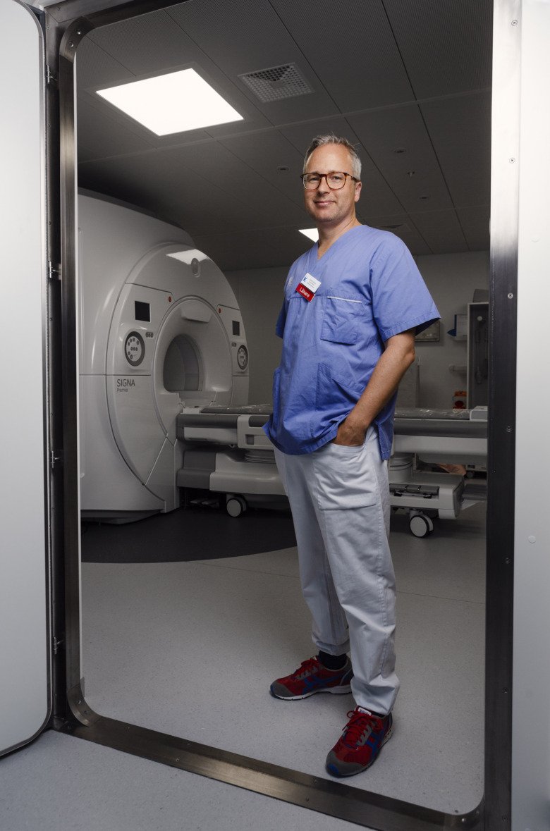
“We currently rely on radiologists to work beyond retirement age, often up until 75 years of age,” says Fredrik Strand, a researcher at Karolinska Institutet’s Department of Oncology-Pathology and himself a radiologist at Karolinska University Hospital.
In his research he studies whether AIs can be used to complement or even replace the role of today’s radiologists in the initial review of mammographs from routine screenings.
In-depth examination
The vast majority of women are positive to having their x-rays evaluated by a computer. A survey conducted by Fredrik Strand’s research group shows that this applies to women across all age groups and especially if AI is permitted to determine the need for an in-depth examination using magnetic resonance imaging (MRI).
But is AI ready to do the job of an experienced radiologist? In a recently published study, Fredrik Strand’s research group evaluated three AI algorithms, demonstrating that the best algorithm was at least as accurate as the average radiologist.
“This is a remarkably impressive result given that the AI algorithm was unable to compare previous images or refer to journal notes regarding possible symptoms, as well as the fact that the majority of the images it trained on were from South Korea and yet it performed so well on our Swedish population,” says Fredrik Strand.
The results will now be followed up in a clinical study at Capio St. Göran's Hospital in Stockholm, where the best of the three algorithms will be put to work as a third reviewer alongside two radiologists. The objective is to assess whether the AI contributes to identifying more cases of breast cancer, as well as whether it is feasible to replace one of the radiologists with the AI.
Find more people with cancer
The study is part of a two-part project, Screen Trust. The second part of study will be conducted at Karolinska University Hospital, with researchers using an AI developed in-house in a collaboration between KTH Royal Institute of Technology and Karolinska Institutet. This phase will further increase precision by selecting women who will be offered a supplementary MRI examination.
Approximately 70% of cancers are detected in those who regularly attend mammography examinations. The remaining 30% are detected due to symptoms, often as a result of self-examination by women. Fredrik Strand emphasises that the point of mammography is to detect a tumour long before the woman herself does so.
“The one third of women who discover their own cancer obtain limited benefit from screening. As things stand, we cannot say with certainty which women will suffer, even if we take account of risk factors such as high breast density,” he says.
The hope is that MRI can contribute to diagnosing more people with cancer; however, an MRI examination is at least five times as costly as a routine mammography examination. All diagnostics also contribute to detecting benign (non-cancerous) changes that do not require further investigation. MRI is currently only offered to healthy women with a hereditary risk of developing breast cancer or specific identified gene mutations.
“By the time the study is complete, our AI will have selected around 1,000 women who we will examine using MRI. The aim is to diagnose more people with cancer at an earlier stage than is currently the case and whether or not the combination of AI and MRI is socioeconomically justifiable largely depends on how many more cancers we find,” says Fredrik Strand.
Same way as a radiologist
In principle, an AI algorithm detects cancer in the same way as a radiologist; it compares each new image with an experience bank of earlier images and identifies deviations in the image similar to those seen previously in women with breast cancer. This is made possible by training the AI algorithm with the aid of large numbers of images.
This method is called deep learning, a form of machine learning that uses neural networks with the ability to interpret images in several stages, much as our own brain does. Deep learning has been proven to work well for image recognition and is becoming more widely available thanks to increasingly powerful computers.
Fredrik Strand is convinced that AI has a role to play in breast cancer screening.
“Tomorrow’s screening will be personalised, with some women having a mammograph and others being chosen for a supplementary examination. This may involve a range of methods; for example, contrast agents together with mammography,” he says.
The selection may be made by a single AI that also sends an immediate message to the majority of women, who instead of waiting weeks for notification can leave the clinic in the knowledge that they are healthy. But isn’t there a risk that an AI will make a wrong diagnosis?
“Absolutely, it will happen that a woman goes home with a negative cancer diagnosis and six months later she finds a lump in her breast, but that already happens today in almost one third of cancer cases – in those with a combination of a certain breast anatomy and certain types of tumour. We need to broaden the discussion on the use of AI, among other things regarding the margins of error we are prepared to accept,” says Fredrik Strand.
Facts: Every image is reviewed by two radiologists
Mammography uses two images of each breast obtained using plain film radiography (x-ray). This results in a greyscale image on which fatty tissue appears as darker and connective tissue lighter. Any tumour also appears as a light deviation, making it difficult to see against a large amount of connective tissue and glands (also known as high breast density).
Each image is reviewed by two radiologists. If either of these identify a suspected tumour, the image is flagged for further discussion involving at least two radiologists who together decide whether or not the woman should be recalled for further examination.
Since 1997, in Sweden all women between the ages of 40 and 74 are offered mammography screening.